
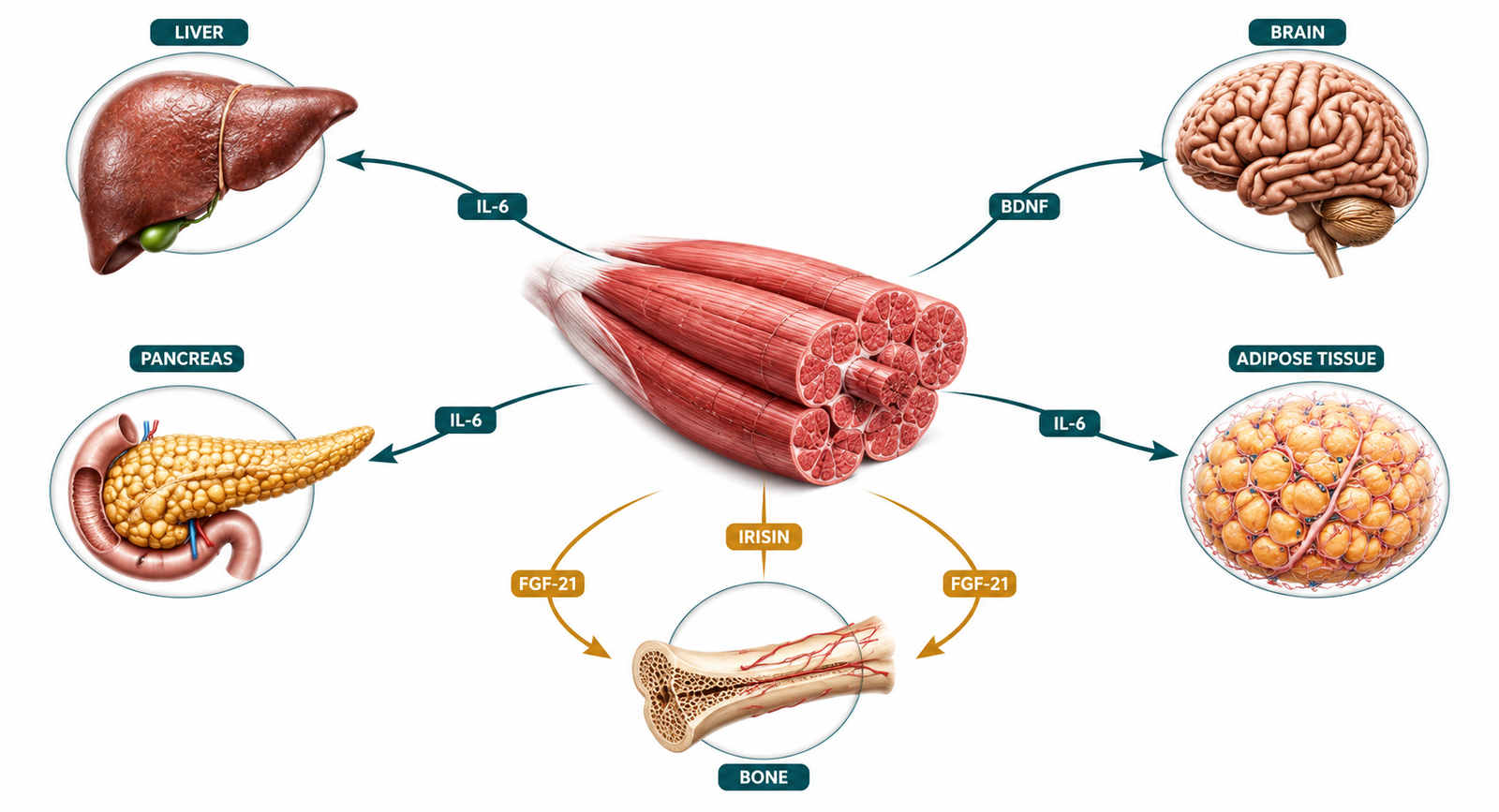
Muscle as an endocrine organ, skeletal muscle does far more than move your body — it broadcasts hormonal signals that regulate inflammation, insulin sensitivity, and metabolic health across every organ system.
When most people think about what muscles do, they think about movement. Lift a weight, walk up a flight of stairs, carry groceries — muscle contracts, force is produced, the job is done. That mechanical picture is accurate but radically incomplete. Over the past two decades, research has established something that has not yet reached mainstream clinical practice: skeletal muscle is a full-spectrum endocrine organ. It produces, secretes, and targets a family of signaling proteins that regulate inflammation, fat metabolism, insulin sensitivity, brain function, and organ crosstalk across the entire body.
The implications of this are significant. When muscle contracts regularly under sufficient mechanical load, it broadcasts a systemic metabolic signal that benefits virtually every other organ. When it does not — when it atrophies from disuse, aging, or chronic underloading — those signals go quiet. The downstream consequences reach far beyond the musculoskeletal system.
This post examines what that endocrine function looks like in practice, which signaling molecules are involved, and why the absence of muscle-derived signals may be one of the most underappreciated drivers of metabolic dysfunction in modern clinical medicine.
What you will learn: What myokines are and how they differ from other hormonal signals | Which specific myokines matter most for metabolic health and why | How the active but metabolically congested phenotype develops | Why exercise signal can be functionally muted even in people who train regularly | What this means practically for reversing insulin resistance
Skeletal Muscle Produces Hormones: The Discovery That Changed the Picture
The endocrine function of skeletal muscle was not always understood. For most of the twentieth century, the prevailing model treated muscle as a target of hormonal signals — insulin, cortisol, testosterone, IGF-1 — rather than a producer of them. That model began to change in the early 2000s when Bente Klarlund Pedersen and colleagues identified interleukin-6 as a cytokine released by contracting skeletal muscle during exercise in concentrations far exceeding what inflammatory sources could account for. The discovery that muscle itself was the source challenged the existing framework and opened a new field of investigation.
The term myokine — derived from the Greek for muscle and movement — was coined to describe cytokines and peptides produced and secreted specifically by skeletal muscle in response to contraction. Since that initial identification, researchers have catalogued over six hundred candidate myokines, with a subset now having well-characterized metabolic effects. The full picture is still being assembled, but what is already established is clinically consequential.
What makes myokines distinct from classical endocrine hormones is their context-dependency. Most traditional hormones are secreted by dedicated glandular tissue in response to a defined stimulus and act on distant target organs through the bloodstream. Myokines are secreted in proportion to mechanical load and contraction frequency, act locally within the muscle, regionally on adjacent fat tissue, and systemically on distant organs including the liver, pancreas, brain, and bone. They are not produced by a dedicated organ — they are produced by the largest organ in the body, and their output is determined by how much that organ is actually being used.
The Key Myokines and What They Do
Interleukin-6: The Anti-Inflammatory Paradox
IL-6 is the most studied myokine and the one most frequently misunderstood in clinical discussions. In the context of obesity, adipose tissue inflammation, and chronic metabolic dysfunction, IL-6 is elevated and pro-inflammatory — a signal of immune activation and metabolic stress. This is the IL-6 that appears on inflammatory panels and that clinicians rightly associate with disease progression.
Muscle-derived IL-6 operates differently. During sustained muscle contraction, IL-6 is released in large quantities from active muscle fibers and acts as a metabolic signal rather than an inflammatory one. It stimulates fat oxidation in working muscle, enhances hepatic glucose production during exercise to maintain fuel availability, and — critically — suppresses the production of TNF-α and IL-1β, two genuinely pro-inflammatory cytokines. Muscle-derived IL-6 is anti-inflammatory. The same molecule has opposing biological effects depending on whether it originates from contracting muscle or from inflamed adipose tissue.
This distinction is not merely academic. In sedentary individuals or those with significant muscle loss, the anti-inflammatory IL-6 signal from muscle is absent. The pro-inflammatory IL-6 from dysfunctional adipose tissue is not. The net cytokine environment shifts toward chronic low-grade inflammation — one of the most consistent upstream drivers of insulin resistance.
Irisin: Fat Browning and Insulin Sensitivity
Irisin is cleaved from a membrane protein called FNDC5 and released predominantly during resistance exercise. Its primary documented action is the promotion of white adipose tissue browning — the conversion of metabolically inert fat storage cells into beige adipocytes that dissipate energy as heat rather than storing it. This thermogenic shift reduces the mass and inflammatory burden of visceral fat, which is itself a major driver of hepatic insulin resistance.
Beyond adipose tissue, irisin has documented effects on bone mineral density, neuronal plasticity, and insulin sensitivity in peripheral tissues. It also appears to mediate some of the cognitive benefits of exercise through its action on BDNF expression in the hippocampus. Sedentary muscle produces very little irisin. Active, mechanically loaded muscle produces it in proportion to the exercise stimulus — particularly resistance training.
BDNF: The Brain-Muscle Connection
Brain-derived neurotrophic factor is produced in the central nervous system but is also secreted by contracting skeletal muscle and crosses the blood-brain barrier. During exercise, circulating BDNF rises substantially — and this increase is now understood to mediate many of the well-established cognitive benefits of physical activity, including improvements in memory, executive function, and mood regulation.
From a metabolic standpoint, BDNF also plays a role in hypothalamic regulation of energy balance and appetite. Its reduction with inactivity and muscle loss may contribute to the dysregulated hunger and reward signaling that complicates metabolic recovery in chronically sedentary patients.
FGF-21, IGF-1, and Myonectin
Fibroblast growth factor 21 (FGF-21) is produced by multiple tissues including liver and adipose, but skeletal muscle also secretes it during exercise and fasting states. Muscle-derived FGF-21 acts locally to enhance fatty acid oxidation and improve mitochondrial function — a function that becomes particularly relevant in the context of metabolic inflexibility, where the ability to shift between glucose and fat as fuel sources is impaired.
IGF-1 produced locally within muscle tissue in response to mechanical loading drives muscle protein synthesis and satellite cell activation — the cellular machinery of muscle repair and growth. Unlike hepatic IGF-1, which is systemically regulated, local muscle IGF-1 is responsive to the mechanical environment. Underload the muscle, and local IGF-1 production drops, impairing the maintenance of muscle mass independent of systemic hormonal status.
Myonectin, a more recently characterized myokine, suppresses autophagy in adipose tissue and promotes fatty acid uptake in the liver and adipose tissue following exercise — contributing to the coordinated post-exercise clearance of circulating lipids.

Clinical Perspective: What I See in Practice
One of the most instructive patterns in my clinical practice involves patients who are genuinely active — they go to the gym, they move — but whose metabolic markers tell a different story. Elevated fasting insulin. A TG/HDL ratio above 2.5. Central fat that resists change despite consistent training. Post-meal fatigue that should not be present in someone who exercises regularly.
What I consistently find is that the exercise signal is being overpowered rather than absent. These patients are producing myokines — their muscles are contracting — but the anti-inflammatory and insulin-sensitizing effects of those signals are being drowned out by a louder metabolic noise: chronically high refined carbohydrate intake that keeps insulin suppressed for fat oxidation around the clock, constant snacking that eliminates the fasting periods in which irisin and FGF-21 exert their metabolic repair functions, poor sleep that elevates cortisol and blunts insulin sensitivity regardless of what happened in the gym, and in many cases a training structure built around high-intensity cardio that serves as an additional cortisol load rather than a clean anabolic signal.
There is also a pattern I see repeatedly in what I call the active sedentary: a person who completes one focused workout per day but remains largely immobile for the remaining twenty-two or twenty-three hours. From a myokine secretion standpoint, this is not equivalent to frequent lower-intensity movement distributed across the day. Total daily contraction time matters, not just peak contraction intensity. A patient who trains hard for forty-five minutes and then sits for eight hours is producing a fraction of the cumulative myokine output of someone who moves moderately but consistently throughout the day.
Another pattern worth naming directly: the distinction between performance muscle and metabolic muscle. Some patients have trained themselves to perform — they are strong, they have capacity — but their muscle tissue has not developed the mitochondrial density or habitual glycogen turnover needed to function as an effective metabolic buffer. High-rep, high-intensity training without adequate recovery and protein support can build a body that looks capable while remaining metabolically underequipped. The muscle is there. The endocrine output is not proportional to its mass.
In practice, when I see an active patient with persistent hyperinsulinemia, I do not tell them to exercise more. I ask what their food pattern looks like between sessions, how their sleep is structured, what their daily step count is outside of formal training, and how much protein they are actually consuming. The exercise is rarely the problem. The twenty-three hours around it almost always are.
Why the Absence of Myokine Signaling Matters Systemically
The myokine framework reframes what muscle loss actually means for metabolic health. Sarcopenia — the progressive loss of muscle mass with aging and disuse — is commonly framed in terms of reduced strength and increased fall risk. Those are real consequences. But the endocrine dimension adds a further layer: as muscle mass and contraction frequency decline, the body loses a major source of anti-inflammatory signaling, insulin-sensitizing hormones, fat browning stimuli, and organ-protective peptides.
The liver is particularly affected. Muscle-derived IL-6 and FGF-21 both modulate hepatic lipid metabolism and glucose output. When those myokine signals are absent, hepatic insulin resistance deepens and de novo lipogenesis — the conversion of carbohydrate substrate into liver fat — accelerates. This is one of the mechanistic links between sarcopenia and non-alcoholic fatty liver disease: the muscle loss is not just a parallel finding, it is a causal upstream driver of liver fat accumulation.
The pancreas is affected as well. Irisin and IL-6 both have documented protective effects on pancreatic beta cell function. As myokine output declines with muscle loss, beta cells lose a significant source of trophic support, potentially accelerating the progression from compensatory hyperinsulinemia to beta cell exhaustion.
The brain is affected through the BDNF pathway. The cognitive decline that frequently accompanies metabolic deterioration — the brain fog, reduced working memory, attentional drift — is not purely a consequence of glucose dysregulation. It also reflects the loss of exercise-derived neurotrophic support that contracting muscle normally provides.
The Endocrine Argument for Resistance Training
Understanding muscle as an endocrine organ reframes resistance training in a way that most patients — and most clinicians — have not encountered. It is not exercise for the sake of burning calories or building aesthetics. It is the activation of the body’s largest hormonal gland.
When a patient with insulin resistance begins a structured resistance training program, the metabolic benefits they experience are not simply the result of increased glucose disposal during the workout. They reflect the systemic hormonal signal that regular muscle contraction broadcasts across every organ system: reduced inflammatory tone via muscle-derived IL-6, improved fat oxidation capacity via irisin and FGF-21, enhanced hepatic lipid handling, better pancreatic function, and improved neurological regulation.
This is also why the type and structure of training matters metabolically. Resistance training under sufficient mechanical load — the kind that produces meaningful muscle protein synthesis and mechanical tension — generates a qualitatively different myokine profile than low-load repetitive movement. Volume and consistency matter across the week, not just peak session intensity. And because myokine signaling is context-dependent, the nutritional and recovery environment around training determines how much of that signal is actually received.

The Animal-Based Protocol for Insulin Resistance and Type 2 Diabetes
A Functional Medicine Framework
A Note on Uncertainty
The myokine field is active and still developing. While the actions of IL-6, irisin, BDNF, and FGF-21 are well-supported by a substantial body of research, many of the over six hundred candidate myokines identified to date have not yet had their mechanisms or clinical significance fully characterized. Some findings from animal studies have not translated cleanly to human physiology, and the dose-response relationships between specific exercise modalities and myokine output are not yet precisely mapped. What is established is the directional evidence: muscle contraction produces systemic anti-inflammatory and insulin-sensitizing signals, and the loss of that signaling has measurable metabolic consequences. The mechanistic details continue to be refined.
People Also Ask
What is a myokine?
A myokine is a cytokine or peptide hormone produced and secreted by skeletal muscle in response to contraction. Unlike classical hormones produced by dedicated glandular tissue, myokines are released in proportion to mechanical load and contraction frequency, and act locally, regionally, and systemically to regulate inflammation, insulin sensitivity, fat metabolism, and organ function.
Does muscle produce hormones?
Yes. Skeletal muscle produces a broad family of signaling proteins called myokines that act on the liver, adipose tissue, brain, pancreas, and bone. This makes muscle a genuine endocrine organ — not merely a target of hormonal signals but an active producer of them.
What does IL-6 from muscle do?
Muscle-derived IL-6, released during sustained contraction, acts as an anti-inflammatory metabolic signal. It stimulates fat oxidation, enhances insulin sensitivity in peripheral tissues, and suppresses the production of pro-inflammatory cytokines including TNF-α. This is distinct from IL-6 produced by inflamed adipose tissue, which acts as a pro-inflammatory signal.
What is irisin and why does it matter?
Irisin is a myokine released predominantly during resistance exercise that promotes the browning of white adipose tissue, improves insulin sensitivity, supports bone mineral density, and appears to mediate some of the cognitive benefits of exercise through its effects on BDNF. Its production is substantially lower in sedentary individuals.
Can you be active and still have insulin resistance?
Yes. Regular exercise does not automatically prevent or reverse insulin resistance if the nutritional environment, sleep quality, and daily movement pattern outside of formal training are suboptimal. The myokine signals produced during exercise can be functionally outweighed by chronic hyperinsulinemia from frequent carbohydrate intake, inflammatory load, and circadian disruption.
Why does muscle loss worsen fatty liver?
Muscle loss reduces the anti-inflammatory and lipid-regulating myokine signals that contracting muscle normally delivers to the liver. It also reduces the body’s total glucose disposal capacity, increasing the substrate available for hepatic de novo lipogenesis. These two mechanisms together make sarcopenia a significant upstream driver of fatty liver progression.
What type of exercise produces the most beneficial myokines?
Resistance training under sufficient mechanical load produces the strongest irisin and IGF-1 response. Sustained moderate-intensity aerobic activity generates the largest IL-6 response. Both modalities produce BDNF and FGF-21. Frequency and consistency of daily movement — not just peak session intensity — determines cumulative myokine output across the day.
About the Author
Morteza Ariana is a State-Certified Functional Nutritionist based in Germany, specializing in insulin resistance, type 2 diabetes, and root-cause metabolic restoration. He holds advanced training in systems-based physiology and has worked with patients across the U.S. and Europe for over 10 years.
His clinical framework is built around a core principle that mainstream medicine consistently overlooks: chronically elevated insulin — not blood glucose — is the earliest and most actionable driver of metabolic disease. That conviction was shaped in part by his own experience with hyperinsulinemia in 2016, and deepened through a decade of clinical practice and the study of leading researchers in metabolic medicine including Benjamin Bikman, Joseph Kraft, Gerald Reaven, Jason Fung, and Stephen Phinney.
His work focuses on identifying and correcting the upstream metabolic signals — insulin load, liver-gut axis dysfunction, circadian misalignment, and micronutrient gaps — that standard screening misses entirely. Patient outcomes are documented, anonymized, and published on this site.
If this resonates, the next step is clarity
The Metabolic Restoration Blueprint is a structured 12-week framework designed to correct upstream metabolic drivers — not just manage symptoms.
Scientific References
Brandt C, Pedersen BK. The role of exercise-induced myokines in muscle homeostasis and the defense against chronic diseases. J Biomed Biotechnol. 2010;2010:520258. 🔗 https://pubmed.ncbi.nlm.nih.gov/20224659/
Pedersen BK, Febbraio MA. Muscles, exercise and obesity: skeletal muscle as a secretory organ. Nat Rev Endocrinol. 2012;8(8):457–465. 🔗 https://pubmed.ncbi.nlm.nih.gov/22473333/
Pedersen BK, Steensberg A, Fischer C, et al. Searching for the exercise factor: is IL-6 a candidate? J Muscle Res Cell Motil. 2003;24(2–3):113–119. 🔗 https://pubmed.ncbi.nlm.nih.gov/14609022/
Boström P, Wu J, Jedrychowski MP, et al. A PGC1-α-dependent myokine that drives brown-fat-like development of white fat and thermogenesis. Nature. 2012;481(7382):463–468. 🔗 https://pubmed.ncbi.nlm.nih.gov/22237023/
Wrann CD, White JP, Salogiannnis J, et al. Exercise induces hippocampal BDNF through a PGC-1α/FNDC5 pathway. Cell Metab. 2013;18(5):649–659. 🔗 https://pubmed.ncbi.nlm.nih.gov/24120943/
Catoire M, Kersten S. The search for exercise factors in humans. FASEB J. 2015;29(5):1615–1628. 🔗 https://pubmed.ncbi.nlm.nih.gov/25609425/
Kim HJ, So B, Choi M, et al. Resistance exercise training increases the expression of irisin concomitant with improvement of muscle function in aging mice and humans. Exp Gerontol. 2015;70:11–17. 🔗 https://pubmed.ncbi.nlm.nih.gov/26169350/
Pedersen BK. Physical activity and muscle-brain crosstalk. Nat Rev Neurosci. 2019;20(7):419–430. 🔗 https://pubmed.ncbi.nlm.nih.gov/31048807/
Iizuka K, Machida T, Hirafuji M. Skeletal muscle is an endocrine organ. J Pharmacol Sci. 2014;125(2):125–131. 🔗 https://pubmed.ncbi.nlm.nih.gov/24837545/
Schnyder S, Handschin C. Skeletal muscle as an endocrine organ: PGC-1α, myokines and exercise. Bone. 2015;80:115–125. 🔗 https://pubmed.ncbi.nlm.nih.gov/26453501/
Severinsen MCK, Pedersen BK. Muscle-organ crosstalk: the emerging roles of myokines. Endocr Rev. 2020;41(4):594–609. 🔗 https://pubmed.ncbi.nlm.nih.gov/32393961/
Febbraio MA, Pedersen BK. Contraction-induced myokine production and release: is skeletal muscle an endocrine organ? Exerc Sport Sci Rev. 2005;33(3):114–119. 🔗 https://pubmed.ncbi.nlm.nih.gov/16006818/
Ahima RS, Park HK. Connecting myokines and metabolism. Endocrinol Metab (Seoul). 2015;30(3):235–245. 🔗 https://pubmed.ncbi.nlm.nih.gov/26354480/
Daskalopoulou SS, Cooke AB, Gomez YH, et al. Plasma irisin levels progressively increase in response to increasing exercise workloads in young, healthy, active subjects. Eur J Endocrinol. 2014;171(3):343–352. 🔗 https://pubmed.ncbi.nlm.nih.gov/24951107/