
Insulin resistance does not begin with high blood sugar. It begins a decade earlier — and it is entirely measurable.
A Systems Biology Perspective
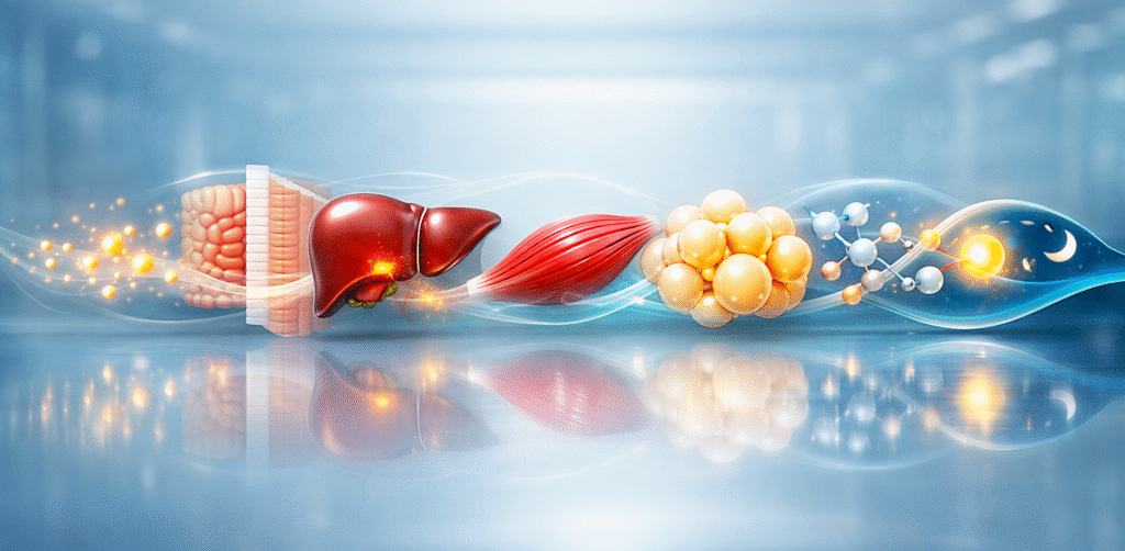
Insulin resistance is a state where your tissues require higher insulin levels to manage the same glucose and energy load. It can develop for years while fasting glucose and HbA1c remain “normal.”
In this guide you’ll learn what insulin resistance is, early signs and lab markers that reveal it, why it progresses, and the principles that make reversal biologically possible.
Medical Disclaimer
Educational information, not medical advice. Work with your clinician, especially if on glucose-lowering meds.
Introduction — The Invisible Beginning
Most metabolic disease does not begin with high blood sugar. It begins years earlier, silently, at the level of cellular signaling and energy regulation.
- A person can have:
- normal fasting glucose
- normal HbA1c
- acceptable LDL numbers
…and still be metabolically dysfunctional.
This early state is often invisible to routine screening, yet it is the phase in which prevention and reversal are most biologically realistic. Insulin resistance is not merely a carbohydrate intolerance or a glucose issue. It is a systemic adaptation to chronic energy surplus, disrupted signaling rhythms, and impaired metabolic flexibility.
To understand it properly, we must move beyond isolated lab markers and view metabolism as an integrated signaling network rather than a calorie equation.
Table of Contents
What Insulin Resistance Actually Is
Insulin resistance is frequently described as “cells not responding to insulin.” While partially correct, this phrasing is incomplete and misleading. Rather than a failure of insulin binding itself, insulin resistance more commonly reflects altered intracellular signaling within a metabolically saturated environment.
Insulin resistance is a state in which tissues require higher levels of insulin to achieve the same metabolic effect, due to altered cellular signaling and energy overflow. This compensatory state — chronically elevated insulin in the bloodstream, known as hyperinsulinemia — is the central driver of the disease cascade and is best assessed through fasting insulin and HOMA-IR rather than glucose alone.
This is not a binary condition.
It exists on a spectrum and can fluctuate over time.
Insulin itself is not the enemy.
It is a storage and signaling hormone that:
- promotes glucose uptake
- regulates amino acid transport
- influences lipid metabolism
- signals growth and repair states
Resistance arises when the signaling environment becomes chronically saturated. The body is not “failing”; it is adapting to prolonged excess and constant stimulation.
Insulin resistance is increasingly understood as a systemic metabolic adaptation involving tissue communication, energy handling, and intracellular signaling, rather than a simple defect in insulin binding, as summarized in major endocrine review literature. Nature Reviews Endocrinology
The causes of insulin resistance are rarely attributable to a single nutrient and more commonly arise from chronic energy surplus, disrupted circadian rhythms, and persistently elevated insulin signaling.
The Energy Overflow Model
A helpful lens is the energy overflow model.
When energy intake chronically exceeds cellular oxidative capacity, substrates accumulate. The liver begins converting excess energy into triglycerides. Adipose tissue expands. Muscle tissue becomes less efficient at glucose uptake. The system shifts from flexible fuel switching to rigid storage bias.
Insulin resistance, in this context, is not random malfunction.
It is a protective buffering response against continuous nutrient exposure.
Key drivers include:
- persistent caloric surplus
- frequent eating with no fasting windows
- circadian disruption
- sleep insufficiency
- low muscle activity
- chronic psychological stress
The problem is rarely one nutrient.
It is the absence of metabolic rhythm.
This concept is discussed further in our article on metabolic flexibility.
The Gut–Liver Axis and Modern Environmental Contributors
Metabolic health is influenced not only by total energy intake and physical activity, but also by the quality of the intestinal environment and the signaling relationship between the gut and the liver — often referred to as the gut–liver axis.
The intestinal barrier functions as a selective interface between the external world and the bloodstream. When this barrier is compromised, small inflammatory molecules and bacterial fragments can translocate into circulation and reach the liver through the portal vein. This process may contribute to low-grade inflammation and altered hepatic insulin signaling.
Several modern lifestyle factors are associated with increased intestinal stress and metabolic disruption:
- high intake of refined carbohydrates and fructose-dense sweeteners
- frequent consumption of ultra-processed foods
- disrupted meal timing and constant snacking
- insufficient dietary protein and micronutrient density
- chronic psychological stress and sleep deprivation
- environmental chemical exposure from plastics, pollutants, and food packaging
- individual sensitivities to certain food components such as gluten or specific plant lectins in susceptible persons
These factors do not affect everyone equally, and metabolic resilience varies between individuals. However, when combined with energy surplus and low physical activity, they may amplify inflammatory signaling and hepatic lipid accumulation, thereby contributing to insulin resistance.
This perspective does not imply that a single nutrient or food group is solely responsible for metabolic dysfunction. Rather, it highlights the cumulative impact of dietary patterns, environmental exposures, and intestinal integrity on liver metabolism and systemic insulin sensitivity.
Restoring gut–liver communication often involves improving food quality, increasing nutrient density, reducing ultra-processed intake, supporting regular meal timing, and addressing sleep and stress patterns. These adjustments work synergistically with muscle activation and fasting windows to restore metabolic balance.
The Liver–Muscle–Adipose Axis
Metabolism is coordinated primarily across three major tissues:
1. The Liver
The liver acts as a metabolic regulator.
It controls glucose release, triglyceride synthesis, and detoxification pathways. When exposed to constant substrate inflow, hepatic fat accumulation increases, often long before symptoms appear.
Elevated triglycerides, rising ALT or GGT, and increasing waist circumference frequently reflect hepatic stress and the insulin-driven fatty liver mechanism rather than dietary fat intake alone.
2. Skeletal Muscle
Muscle is the largest glucose sink in the human body.
Insufficient muscle activation reduces glucose clearance efficiency. Resistance training and movement improve insulin sensitivity not by “burning calories” but by enhancing glucose transporters and mitochondrial density.
3. Adipose Tissue
Adipose tissue is not inert storage. It is an endocrine organ that secretes signaling molecules influencing inflammation and insulin sensitivity. When storage capacity is exceeded — particularly in the visceral compartment versus the subcutaneous compartment — inflammatory signaling increases and insulin resistance accelerates.
These tissues operate as a network.
Disruption in one influence the others.
Early Signs and Tests for Insulin Resistance (Beyond Glucose)
Glucose is a late marker.
Earlier signals often appear in lipid ratios and fasting insulin levels.
Important contextual markers include:
- Triglyceride to HDL ratio — the most under-utilized cardiometabolic ratio in conventional practice
- Fasting insulin concentration — the marker that reveals compensation years before HbA1c shifts, interpreted against the fasting insulin optimal range that reflects metabolic health rather than population averages
- Waist-to-height ratio — visceral adiposity is mechanistically distinct from total body fat
- ALT and GGT trends — early hepatic stress signals
- High-sensitivity CRP context — chronic low-grade inflammation tone
These markers reflect metabolic strain before diabetes manifests.
They reveal direction, not merely status. A normal HbA1c does not guarantee metabolic health.
It simply indicates average glucose exposure, not signaling quality.
The Phenotype Spectrum
Insulin resistance does not always present with obesity.
Several phenotypes exist:
Lean Insulin Resistance
Individuals may appear thin yet exhibit high fasting insulin, elevated triglycerides, or hepatic fat accumulation. This pattern — characterized by insulin resistance with normal blood sugar in the compensatory phase — is linked to chronic stress, poor sleep, or high refined carbohydrate intake combined with low muscle mass.
Athletic Dysregulation
Endurance athletes with excessive training volume but insufficient recovery can develop paradoxical metabolic rigidity and hormonal disruption. The full picture of insulin resistance in athletes is its own clinical entity.
“Normal Labs” Illusion
Many individuals remain within laboratory reference ranges while trending toward dysfunction. Reference ranges reflect population averages, not optimal physiology. Metabolic disease is often gradual and silent.
Waiting for overt pathology delays intervention.
Clinical Perspective: What I See in Practice
The overarching pattern I see across insulin resistance patients in clinical practice is metabolic overcompensation. Patients arrive with a recognizable constellation of symptoms: persistent fatigue, systemic inflammation, central adiposity, and insatiable hunger despite what they describe as adequate caloric intake. They often rely heavily on caffeine to function. Sleep quality is poor. They are frustrated, having been previously reassured that their laboratory results were “not bad enough yet.”
When I run a deeper panel, a consistent laboratory signature emerges. Fasting insulin is elevated, often years before any dramatic glucose abnormality appears. The triglyceride-to-HDL ratio is suboptimal. Waist circumference is increased. ALT and GGT are mildly elevated more often than not. HbA1c shows a creeping upward trend even when it has not yet crossed a diagnostic threshold. Inflammatory markers like hsCRP are frequently elevated. Vitamin D and magnesium deficiencies are common. Uric acid presents in the high-normal or elevated range, reflecting the metabolic burden of high-fructose corn syrup, suboptimal electrolyte status, and the systemic impact of chronic hyperinsulinemia.
What conventional medicine consistently overlooks is this hyperinsulinemic compensation phase. The prevailing approach waits for overt glucose dysregulation, failing to recognize that these patients are not “fine” — their bodies have been maintaining euglycemia for years by producing excessive amounts of insulin to compensate for declining sensitivity. By the time glucose crosses a diagnostic threshold, that compensation has been operating, and damaging the system, for a decade or longer.
The opening conversation I have with these patients is direct but empathetic. Your body is not broken. It is adapting to an environment that is asking too much of your insulin system. That single reframe — from disease identity to biological adaptation — is often what allows the rest of the clinical work to begin. Patients who have been told for years that they have a chronic, progressive condition need to hear that the condition is, in many cases, a reversible adaptation.
The diagnostic work then identifies the primary drivers contributing to the metabolic state: high refined carbohydrate load, frequent eating without metabolic rest, visceral fat and fatty liver, poor sleep hygiene, insufficient muscle mass, chronic stress physiology, and circadian rhythm disruption. The intervention addresses these drivers in a sequence calibrated to the patient.
When treatment is initiated correctly, the initial improvements are often not what patients expect. Weight loss is rarely the first sign of restoration. Patients first report a quieting of hunger and a significant reduction in cravings. Fasting glucose stabilizes. Energy becomes sustained throughout the day rather than collapsing at 2pm. Sleep deepens. Waist circumference reduces noticeably. Systemic inflammation and the puffiness associated with it visibly decrease. Crucially, the patient’s confidence in their body’s ability to heal begins to return — and that subjective shift often precedes the lab improvements that come over the following weeks. Clinically, the focus shifts profoundly from glucose management to the more fundamental goal of restoring insulin sensitivity and metabolic flexibility.
Several patterns are worth naming for clinicians who treat this population. Many patients arrive saying “I don’t even eat that much,” and they are usually telling the truth. By the time they seek help, their metabolism has already downregulated significantly. The core issue is not their current caloric intake — it is the cumulative effect of years of adverse metabolic signaling compounded by low muscle mass and chronic circadian disruption.
A frequent patient question is “why is my glucose high in the morning when I didn’t eat?” The answer is the convergence of elevated hepatic glucose output, disruption of the normal cortisol rhythm, and insulin resistance driven by fatty liver. This phenomenon is often the earliest visible sign of hepatic dysfunction rather than an unexplained anomaly.
The most common patient misunderstanding is that glucose itself is the problem. Clinically, the true issue revolves around insulin dynamics, liver health, and overall energy handling. Glucose is a downstream marker of these deeper systemic imbalances, not the disease itself.
The lab pattern that conventional screening overlooks most consistently is what I call the silent progression phase of metabolic dysfunction: normal or only mildly elevated HbA1c paired with high fasting insulin, a poor triglyceride-to-HDL ratio, a slightly elevated GGT, and high-normal uric acid. This constellation can persist for ten to twenty years without triggering a diagnosis under conventional screening protocols. Patients are repeatedly reassured that “everything is fine” until glucose unequivocally crosses the diagnostic threshold. By then, intervention has come far too late.
My treatment principle follows from this: lower the need for insulin before attempting to improve glucose. This means reducing glycemic load, decreasing eating frequency, actively increasing muscle mass, optimizing sleep and circadian timing, and reducing hepatic fat. Glucose normalizes when the underlying insulin pressure is removed. It does not normalize durably when the insulin pressure is left in place.
Finally, the gut-liver axis is a massively underestimated component in a significant subset of these patients. Microbiome dysbiosis, increased intestinal permeability, LPS translocation, and chronic low-grade inflammation contribute directly to hepatic insulin resistance. These patients often present with bloating, IBS-type symptoms, elevated GGT and ALT, and stubborn insulin resistance despite adhering to a clean diet. In these individuals, the gut-liver axis is not a secondary issue but a primary amplifier of their metabolic dysfunction.
The bottom-line clinical observation is this: most insulin resistance is not a singular problem. It is a network failure involving the liver, muscle, adipose tissue, gut, and brain — particularly in appetite and circadian control. Treating individual nodes can offer some benefit, but true restoration of the entire system is what ultimately changes the patient’s metabolic trajectory.
Signaling Rhythms — mTOR and AMPK
Metabolism operates through oscillating signaling states.
Growth and Repair (mTOR Dominance)
Activated during feeding, protein intake, and resistance training. Supports tissue growth, immune function, and cellular repair.
Maintenance and Cleanup (AMPK Dominance)
Activated during fasting, exercise, and energy scarcity. Supports mitochondrial biogenesis, autophagy, and metabolic efficiency.
Health is not about choosing one pathway permanently.
It is about rhythmic alternation.
Constant feeding, constant snacking, and continuous stimulation suppress AMPK periods and reduce metabolic flexibility. Conversely, excessive restriction without nourishment impairs repair pathways.
Balance emerges from structured cycles, not extremes.
Further reading on protein and mTOR signaling pathway
Circadian Influence and Light Exposure
Metabolism is tightly linked to circadian biology. Light exposure influences hormonal signaling, sleep quality, and insulin sensitivity through pathways that operate independently of caloric intake or physical activity. The body’s metabolic timing system expects specific light, food, and rest signals at specific points in the 24-hour cycle, and when those signals become misaligned, the metabolic system progressively loses its ability to regulate energy correctly.
Disruption of this system occurs through several recognizable patterns. Artificial light extending wake periods past the biological evening delays melatonin onset and impairs the overnight metabolic recovery that should occur during sleep. Sleep that is shortened or fragmented prevents the hormonal and cellular repair processes that depend on adequate sleep architecture. Meal timing that conflicts with natural rhythms — late-night eating, frequent grazing, or consumption during the biological night — places food into a metabolic context where insulin secretion is suppressed, glucose tolerance is reduced, and hepatic glucose handling is dysregulated.
The clinical consequence is that late-night eating and chronic blue-light exposure can impair glucose regulation independently of total caloric intake. Two patients eating the same diet on different timing schedules will not produce the same metabolic results. Metabolic health is not solely nutritional — it is also temporal.
For the full clinical framework on how sleep and the body clock together govern metabolic health, see the cluster cornerstone on sleep and circadian rhythm.
Consequence Trajectory
When insulin resistance is left unaddressed, the metabolic strain progresses along a recognizable trajectory of downstream consequences. The earliest manifestation is typically fatty liver disease, as the liver accumulates triglycerides in response to chronic insulin and substrate overload. Hypertriglyceridemia follows, reflecting the liver’s increased VLDL output and the system’s reduced capacity to clear circulating lipids.
Hypertension emerges as vascular insulin signaling deteriorates and sodium handling shifts. Type 2 diabetes appears when pancreatic beta cells can no longer produce enough insulin to overcome the accumulated peripheral resistance. Chronic systemic inflammation builds across all of these stages, accelerating each one. Cognitive decline, including the brain insulin resistance now recognized as a contributor to Alzheimer’s disease, develops over decades as the central nervous system loses access to the metabolic infrastructure it depends on.
These are not isolated diseases. They are sequential manifestations of prolonged metabolic strain, all driven by the same upstream dysfunction. Recognizing this trajectory early — at the hyperinsulinemic phase, before any single endpoint disease has crystallized — is what allows redirection before irreversible damage occurs.
How to Reverse Insulin Resistance (Principles That Work)
Reversal is biologically realistic because insulin resistance, in its early and middle phases, is an adaptive state rather than a degenerative one. The body is not failing — it is responding to chronic conditions, and when those conditions change, the response changes with them. Restoration is therefore not about suppressing individual pathways or imposing artificial constraints on the body. It is about returning the system to the rhythm, capacity, and signaling balance that allow it to regulate energy on its own.
The first principle is muscle activation. Skeletal muscle is the largest insulin-sensitive tissue in the body and the primary site of glucose disposal. Resistance training improves glucose transport at the cellular level, increases mitochondrial density, and enhances the muscle’s capacity to clear glucose without requiring large insulin signals. Even modest improvements in muscle mass and training consistency produce measurable metabolic benefits, often within weeks. The mechanism is not caloric expenditure during the training session itself — it is the structural and metabolic upgrade of the tissue that follows.
The second principle is structured fasting windows. Periods without caloric intake allow AMPK activation, lipid mobilization, and the metabolic flexibility that chronic feeding suppresses. The intervention is not extreme fasting or rigid protocols. It is the simple restoration of biological rest — the metabolic quiet between meals that the body was designed for and that modern eating patterns have largely eliminated.
The third principle is protein sufficiency. Adequate amino acid intake supports muscle maintenance, satiety regulation, and the repair processes that hold the metabolic system together. Without sufficient protein, training cannot produce its expected adaptations, satiety signaling becomes unreliable, and the body progressively loses the lean tissue that protects against insulin resistance in the first place.
The fourth principle is sleep and circadian alignment. Consistent sleep duration, fixed wake time, and morning light exposure recalibrate the circadian signaling that governs cortisol rhythm, insulin sensitivity, and appetite regulation. As detailed in the cluster cornerstone referenced earlier, this layer is often the rate-limiting factor in patients whose dietary work has plateaued.
The fifth principle is meal timing awareness. Reducing late-night eating, anchoring the largest meal earlier in the day, and avoiding the chronic grazing pattern that defines modern eating habits all improve glucose regulation, hormonal balance, and the body’s ability to enter the recovery state during sleep.
These five principles share a common quality. They are not extreme interventions. They are not weight-loss tricks or biohacks. They are biological alignments — the structural conditions under which the human metabolic system operates correctly. Restoration emerges from the alignment, not from any single intervention performed in isolation.
For readers who want a structured eating framework that translates these principles into a concrete daily practice, see The Animal-Based Protocol for Insulin Resistance →.
Who Is at Risk for Insulin Resistance?
The framework laid out in this cornerstone is especially relevant for several patient profiles that conventional screening tends to miss.
Individuals with rising triglycerides despite eating what they consider a healthy diet are often in the silent progression phase — the dietary pattern they understand as healthy may be exactly what is driving the metabolic dysfunction, particularly when refined carbohydrates and frequent eating are involved. Professionals experiencing persistent fatigue with normal glucose labs are another classic presentation; the fatigue is often the first symptom of compensatory hyperinsulinemia, appearing years before any glucose marker turns abnormal.
Athletes with declining recovery, paradoxically, can develop the metabolic rigidity described in the insulin resistance in athletes section above, where excessive training volume without adequate recovery produces hormonal and metabolic dysregulation that mimics sedentary insulin resistance through a different mechanism.
Adults noticing an increasing waist circumference without overall weight gain are seeing visceral fat accumulation, which is mechanistically distinct from total body fat and is the strongest predictor of metabolic disease progression. And those with a family history of metabolic disease — type 2 diabetes, fatty liver, cardiovascular disease, or PCOS — carry a predisposition that benefits enormously from earlier identification and intervention rather than waiting for the diagnostic threshold to be crossed.
Recognizing the early symptoms of insulin resistance in any of these profiles allows strategic correction long before standard screening would identify a problem. The absence of overt disease does not equal metabolic health.
Closing Perspective
Insulin resistance is not a moral failure, nor is it an inevitable consequence of aging. It is a physiological adaptation to environmental and behavioral patterns that can be modified.
When metabolism is viewed as a signaling network rather than a calorie ledger, clarity emerges. The goal is not restriction or obsession, but rhythm, capacity, and flexibility.
Understanding these principles transforms metabolic health from a reactive process into a proactive one — shifting the focus from disease management to physiological stewardship.
This manuscript is not a final statement.
It is a living framework, intended to evolve as understanding deepens and evidence expands.
People Also Ask
What are the early symptoms of insulin resistance?
Early insulin resistance symptoms are often silent. When symptoms appear, they may include fatigue after meals, difficulty losing fat despite effort, increased hunger or cravings, brain fog, afternoon energy crashes, and subtle weight gain around the abdomen. These signs reflect altered energy handling and signaling long before blood glucose becomes abnormal.
Can you have insulin resistance with normal blood sugar or HbA1c?
Yes. Many individuals have normal fasting glucose and HbA1c while insulin resistance is already present. In early stages, the body compensates by producing more insulin to keep glucose normal. This compensatory phase can last for years, which is why glucose-based screening alone often misses early metabolic dysfunction.
What is the best test for insulin resistance?
There is no single perfect test. Fasting insulin, HOMA-IR, triglyceride-to-HDL ratio, and oral glucose tolerance tests with insulin measurements each provide partial insight. In practice, insulin resistance is best assessed using patterns across multiple markers combined with clinical context, rather than relying on a single laboratory value.
What is a good fasting insulin or HOMA-IR level?
Rather than strict cutoffs, lower fasting insulin and HOMA-IR values generally reflect better insulin sensitivity. Many metabolically healthy individuals fall toward the lower end of reference ranges. However, interpretation should consider lifestyle, body composition, inflammation, and metabolic flexibility rather than treating any single number as a diagnosis.
How long does it take to improve insulin resistance?
Improvement can begin within weeks, especially with changes in diet quality, meal timing, physical activity, sleep, and stress regulation. However, meaningful and durable restoration of insulin sensitivity often unfolds over months. The timeline depends on baseline metabolic health, consistency of interventions, and removal of chronic stressors driving energy overload.
Is insulin resistance reversible?
In many cases, yes. Insulin resistance is often a functional adaptation, not permanent damage. By restoring metabolic rhythm, reducing chronic energy surplus, improving muscle activity, and supporting circadian alignment, insulin sensitivity can improve substantially. Earlier stages are generally more reversible, but meaningful improvement is possible even after years of dysfunction.
About the Author
Morteza Ariana is a State-Certified Functional Nutritionist based in Germany, specializing in insulin resistance, type 2 diabetes, and root-cause metabolic restoration. He holds advanced training in systems-based physiology and has worked with patients across the U.S. and Europe for over 10 years.
His clinical framework is built around a core principle that mainstream medicine consistently overlooks: chronically elevated insulin — not blood glucose — is the earliest and most actionable driver of metabolic disease. That conviction was shaped in part by his own experience with hyperinsulinemia in 2016, and deepened through a decade of clinical practice and the study of leading researchers in metabolic medicine including Benjamin Bikman, Joseph Kraft, Gerald Reaven, Jason Fung, and Stephen Phinney.
His work focuses on identifying and correcting the upstream metabolic signals — insulin load, liver-gut axis dysfunction, circadian misalignment, and micronutrient gaps — that standard screening misses entirely. Patient outcomes are documented, anonymized, and published on this site.
If this resonates, the next step is clarity
The Metabolic Restoration Blueprint is a structured 12-week framework designed to correct upstream metabolic drivers — not just manage symptoms.
Key References
DeFronzo RA, Tripathy D. Skeletal muscle insulin resistance is the primary defect in type 2 diabetes. Diabetes Care. 2009;32 Suppl 2:S157–S163. 🔗 https://pubmed.ncbi.nlm.nih.gov/19875544/
Reaven GM. Banting lecture 1988. Role of insulin resistance in human disease. Diabetes. 1988;37(12):1595–1607. 🔗 https://pubmed.ncbi.nlm.nih.gov/3056758/
Kraft JR. Detection of diabetes mellitus in situ (occult diabetes). Laboratory Medicine. 1975;6(2):10–22. 🔗 https://academic.oup.com/labmed/article-abstract/6/2/10/2657843
Kahn SE, Hull RL, Utzschneider KM. Mechanisms linking obesity to insulin resistance and type 2 diabetes. Nature. 2006;444(7121):840–846. 🔗 https://pubmed.ncbi.nlm.nih.gov/17167471/
Petersen MC, Shulman GI. Mechanisms of insulin action and insulin resistance. Physiological Reviews. 2018;98(4):2133–2223. 🔗 https://pubmed.ncbi.nlm.nih.gov/30067154/
Samuel VT, Shulman GI. Mechanisms for insulin resistance: common threads and missing links. Cell. 2012;148(5):852–871. 🔗 https://pubmed.ncbi.nlm.nih.gov/22385956/
Czech MP. Insulin action and resistance in obesity and type 2 diabetes. Nature Medicine. 2017;23(7):804–814. 🔗 https://pubmed.ncbi.nlm.nih.gov/28697184/
Crofts CAP, Schofield G, Zinn C, Wheldon M, Kraft J. Identifying hyperinsulinaemia in the absence of impaired glucose tolerance: an examination of the Kraft database. Diabetes Research and Clinical Practice. 2016;118:50–57. 🔗 https://pubmed.ncbi.nlm.nih.gov/27344544/
Bikman BT, Shimy KJ, Apovian CM. A review of the effects of dietary carbohydrate restriction on cardiometabolic markers. Nutrients. 2020;12(10):2954. 🔗 https://pubmed.ncbi.nlm.nih.gov/32987684/
Saklayen MG. The global epidemic of the metabolic syndrome. Current Hypertension Reports. 2018;20(2):12. 🔗 https://pubmed.ncbi.nlm.nih.gov/29480368/
Roden M, Shulman GI. The integrative biology of type 2 diabetes. Nature. 2019;576(7785):51–60. 🔗 https://pubmed.ncbi.nlm.nih.gov/31802013/
Hatting M, Tavares CDJ, Sharabi K, Rines AK, Puigserver P. Insulin regulation of gluconeogenesis. Annals of the New York Academy of Sciences. 2018;1411(1):21–35. 🔗 https://pubmed.ncbi.nlm.nih.gov/28868790/
Lustig RH, Mulligan K, Noworolski SM, et al. Isocaloric fructose restriction and metabolic improvement in children with obesity and metabolic syndrome. Obesity. 2016;24(2):453–460. 🔗 https://pubmed.ncbi.nlm.nih.gov/26499447/
Stenvers DJ, Scheer FAJL, Schrauwen P, la Fleur SE, Kalsbeek A. Circadian clocks and insulin resistance. Nature Reviews Endocrinology. 2019;15(2):75–89. 🔗 https://pubmed.ncbi.nlm.nih.gov/30531917/
Younossi ZM, Golabi P, Paik JM, et al. The global epidemiology of nonalcoholic fatty liver disease (NAFLD) and nonalcoholic steatohepatitis (NASH): a systematic review. Hepatology. 2023;77(4):1335–1347. 🔗 https://pubmed.ncbi.nlm.nih.gov/36626630/
Cani PD, Amar J, Iglesias MA, et al. Metabolic endotoxemia initiates obesity and insulin resistance. Diabetes. 2007;56(7):1761–1772. 🔗 https://pubmed.ncbi.nlm.nih.gov/17456850/
Last reviewed: May 2026. Content by Morteza Ariana, State-Certified Functional Nutritionist, Germany.