
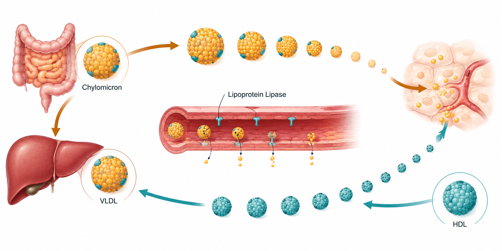
Lipoprotein particle transport: cholesterol and fat move outward via VLDL and chylomicrons, and back to the liver via HDL’s reverse transport route.
A standard lipid panel reads like a set of static measurements — LDL, HDL, triglycerides, each a number sitting still on a page. What it’s actually describing is closer to a shipping network: particles built in the liver and gut, sent into circulation, unloaded piece by piece, remodeled mid-transit, and eventually either recycled or cleared. The individual numbers on a lab report are single frames pulled from a process in constant motion. Understanding lipoprotein particle transport — the whole route cholesterol and fat travel through the bloodstream — is what turns those static numbers into a coherent picture of what’s actually happening.
This post connects the two particle types already covered in this series — LDL and VLDL — into the larger transport system they’re part of: how fat leaves the gut, how the liver exports and later reclaims cholesterol, and how HDL runs the return route in the opposite direction.
What You Will Learn
- The two separate delivery routes that bring fat into circulation — from the gut and from the liver
- How lipoprotein lipase unloads triglyceride from particles at the capillary wall
- What a “remnant” particle is, and why apoE is the key that lets the liver reclaim it
- How HDL runs cholesterol transport in the opposite direction, back to the liver
- Why some particle exchanges make LDL more dangerous, and how CETP drives that process
- Why a full panel read as a transport system reveals more than any single marker in isolation
Two Roads In: The Exogenous and Endogenous Pathways
Fat enters the bloodstream by two separate routes, and it’s worth keeping them distinct because they answer different questions about a patient.
The exogenous pathway starts in the gut. After a meal, the intestine packages dietary fat and cholesterol into chylomicrons — the largest, most triglyceride-rich lipoprotein particles in circulation. Chylomicrons are built to do one job: move a large fat load from a single meal out into the body quickly. This pathway reflects what’s coming in from outside, meal by meal.
The endogenous pathway starts in the liver, and it’s the one already covered in detail in the VLDL metabolism post: the liver packages its own triglyceride supply — whether that fat arrived from adipose tissue or was synthesized internally — into VLDL and exports it. This pathway reflects what the liver itself is doing, independent of any single meal.
Both pathways converge on the same downstream machinery. Chylomicrons and VLDL are structurally similar — both large, triglyceride-rich, both anchored by an apolipoprotein (apoB-48 for chylomicrons, apoB-100 for VLDL) — and both get processed by the same enzyme once they reach the capillary bed.
The Delivery Truck: Lipoprotein Lipase and the Capillary Handoff
Both chylomicrons and VLDL get unloaded at the same site: the luminal surface of capillaries in muscle, heart, and adipose tissue, where an enzyme called lipoprotein lipase (LPL) sits waiting.
LPL doesn’t float freely in the blood. It’s manufactured inside muscle and fat cells, then transported to the capillary surface and anchored there by a specialized protein called GPIHBP1, which acts as a docking platform — shuttling LPL from the tissue side of the capillary wall to the blood-facing surface, where it can actually reach passing particles. Without GPIHBP1, LPL never makes it to where the particles are; this isn’t a minor detail, since GPIHBP1-deficient animals develop severe triglyceride buildup in the blood despite having functional LPL sitting uselessly in the wrong location.
Once anchored, LPL hydrolyzes the triglyceride core of a passing chylomicron or VLDL particle, releasing fatty acids that the adjacent muscle or fat cell absorbs directly for fuel or storage. As the particle’s triglyceride core empties, it shrinks and its relative cholesterol content rises. This is the transition point covered from the LDL side in the earlier post in this cluster: VLDL, stripped of triglyceride, becomes a remnant particle, then eventually LDL. Chylomicrons undergo the same shrinking process, becoming chylomicron remnants.

The Second Address Label: ApoE and Remnant Clearance
VLDL and chylomicron remnants face a choice most particles never encounter: get cleared by the liver quickly, or continue through the delipidation cascade toward LDL.
This choice depends heavily on a second apolipoprotein, apoE, which sits on the remnant surface alongside apoB. Where apoB-100 is the key that unlocks the LDL receptor (covered in the earlier LDL post), apoE is a broader, higher-affinity key — it binds not just the LDL receptor but a whole family of related receptors, plus heparan sulfate proteoglycans that trap remnants in the space between liver cells even before receptor binding occurs. The clearance process runs in stages: proteoglycans in the liver’s sinusoidal space capture remnant particles first, hepatic lipase further processes them there, and receptor-mediated endocytosis finishes the job.
The clinical significance is direct: a remnant particle with plenty of functional apoE tends to get pulled out of circulation quickly by the liver, before it can be extensively remodeled into small, dense LDL. A remnant that lacks functional apoE — as happens in certain genetic apoE variants — lingers in circulation far longer, continues accumulating cholesteryl ester, and becomes a cholesterol-enriched remnant particle that is itself directly atherogenic, independent of anything happening downstream in the LDL pathway. This is why remnant cholesterol has emerged as its own risk marker distinct from LDL cholesterol: a particle that never even reaches the LDL stage can still be doing damage.
The Return Route: HDL and Reverse Cholesterol Transport
Every pathway so far moves cholesterol outward from the liver and gut into peripheral tissue. HDL runs the opposite direction — it’s the return route, pulling excess cholesterol out of peripheral cells (including, critically, macrophages sitting in the artery wall) and carrying it back to the liver for disposal. This return route is called reverse cholesterol transport, and it’s the mechanistic reason HDL has historically been considered protective.
The first step is the one that actually matters clinically: a macrophage that has accumulated cholesterol — the same kind of macrophage building toward a foam cell in the retention process covered in the LDL post — can hand that cholesterol off to a nearby HDL particle, provided the HDL particle is functionally capable of accepting it. This capacity is called cholesterol efflux capacity, and it turns out to be a more accurate predictor of cardiovascular risk than HDL cholesterol concentration itself.
In a large clinical cohort, cholesterol efflux capacity predicted cardiovascular events independent of, and more strongly than, the plain HDL-C number — a finding that directly explains why raising HDL cholesterol pharmacologically has repeatedly failed to reduce cardiovascular events in trials. The quantity of HDL circulating and the functional capacity of that HDL to actually do its job are not the same thing, and only one of them is what a routine panel measures.
Where the Two Directions Meet: CETP and the Traffic Between Particles
The outward and return pathways aren’t independent systems running in parallel — they trade cargo directly, through an enzyme called cholesteryl ester transfer protein (CETP). CETP moves cholesteryl ester out of HDL and into LDL and VLDL, while moving triglyceride in the opposite direction, into HDL.
Under normal conditions this exchange is modest. But when plasma triglycerides are elevated — the exact hepatic overproduction pattern described in the VLDL post — CETP activity intensifies. More cholesteryl ester leaves HDL, more triglyceride arrives in its place, and the triglyceride-loaded HDL becomes a substrate for further enzymatic processing that shrinks it and accelerates its clearance from circulation. This is a major reason elevated triglycerides and low HDL travel together so reliably: they’re not two separate abnormalities, they’re two visible symptoms of the same CETP-driven exchange, running on the same excess triglyceride supply. The identical exchange reshapes LDL on the other side, contributing to the small, dense LDL particle formation already covered in this cluster.
Clinical Perspective: What I See in Practice
When I look at a full lipid panel, I don’t treat LDL-C, triglycerides, HDL-C, and ApoB as separate numbers. I think about lipoprotein traffic: where the particles are coming from, how many are circulating, what they are carrying, how they are being remodeled, and how efficiently they are being cleared.
A classic pattern is relatively normal LDL-C, normal or only mildly elevated triglycerides, reasonable HDL-C, but disproportionately elevated ApoB. That discordance matters because LDL-C tells us how much cholesterol is being carried inside the LDL compartment, whereas ApoB approximates the number of circulating atherogenic particles. Two patients can therefore have the same LDL-C but very different numbers of particles carrying that cholesterol — like two roads transporting the same amount of cargo, but one using 100 large trucks and the other 160 smaller trucks. In metabolic dysfunction, particularly when triglycerides are elevated, particles can become relatively cholesterol-depleted, so LDL-C may look reassuring while the number of ApoB-containing particles remains higher than expected.
I also look upstream at the VLDL → IDL/remnant → LDL pathway. The liver packages triglyceride and cholesterol into VLDL, and as these particles lose triglyceride through lipoprotein lipase and other remodeling processes, they become IDL/remnants and eventually LDL. So a pattern of higher triglycerides, lower HDL-C, relatively ordinary LDL-C, and elevated ApoB makes me think about increased triglyceride-rich lipoprotein traffic, hepatic VLDL production and remodeling, and a greater number of atherogenic particles — not simply “the LDL is fine.” That’s why triglycerides are more informative when interpreted alongside ApoB and non-HDL-C rather than in isolation. I also consider Lp(a) separately, because it is a genetically determined ApoB-containing particle.
What I tell the patient is: your cholesterol isn’t just sitting in your blood as one substance. Your liver packages lipids into transport particles, sends them into circulation, remodels them, and eventually clears them. LDL-C tells us the amount of cholesterol being carried in LDL; ApoB gives us another piece of the picture — the number of atherogenic particles carrying that cholesterol; triglycerides tell us something about the triglyceride-rich traffic; and non-HDL-C captures the cholesterol carried by the broader ApoB-containing pool. If those numbers disagree, I pay attention to the traffic rather than assuming one number tells the whole story.
And if triglycerides, fasting insulin, glucose regulation, liver markers, and ApoB together suggest the liver is actively overproducing and remodeling lipoproteins, I want to understand why that traffic is happening. I’m not interested in treating a number without understanding the system generating it.
In other words, the deeper question is not simply “how much cholesterol do you have?” but “what is your lipoprotein transport system doing with it?”
Clinical Implications
Reading a panel as a transport system rather than a set of isolated numbers changes what gets ordered and what gets explained to a patient.
The first implication is when discordance should trigger additional testing. Elevated triglycerides alongside low HDL and unremarkable LDL-C is precisely the signature of active CETP-driven exchange described above — it’s a reasonable trigger for ordering ApoB or a particle-number test, rather than reassurance from the LDL-C value alone.
The second is that HDL cholesterol, taken alone, is a genuinely limited marker. A low HDL-C in the setting of elevated triglycerides usually reflects the same upstream process driving the rest of the picture — it isn’t an independent deficiency to be corrected in isolation, and raising it pharmacologically without addressing the triglyceride-driving process behind it has not translated into reduced cardiovascular events in trials.
The third is that remnant particles deserve attention on their own terms, not only as a waypoint toward LDL. A patient with elevated remnant cholesterol or a family history suggesting impaired apoE function can carry meaningful atherogenic risk even when LDL-C itself looks unremarkable.
The fourth is sequencing, echoing the same principle from the earlier posts in this cluster: because so much of this traffic pattern is driven by hepatic triglyceride output, addressing insulin resistance and hepatic fat tends to improve the entire transport picture — triglycerides, HDL, and particle number together — rather than requiring separate interventions for each marker.
A Note on Uncertainty
The core transport steps described here — the two entry pathways, LPL-mediated triglyceride hydrolysis, apoE-mediated remnant clearance, and CETP-mediated lipid exchange — are supported by consistent mechanistic and genetic evidence built up over several decades.
Less settled is exactly how much independent risk remnant particles carry once ApoB and LDL particle number are both accounted for, since remnant cholesterol and ApoB are correlated and separating their individual contributions in any one patient isn’t something routine testing resolves cleanly. Similarly, while cholesterol efflux capacity outperforms HDL-C as a predictor in research cohorts, it isn’t yet a standard, widely available clinical test, so in practice most patients are still assessed with the less precise measure. I’ve noted where the evidence is strong and where it’s still developing rather than presenting a tidier picture than currently exists.

The Animal-Based Protocol for Insulin Resistance and Type 2 Diabetes
A Functional Medicine Framework
People Also Ask
What is the difference between chylomicrons and VLDL?
Both are large, triglyceride-rich particles that deliver fat into circulation, but chylomicrons are made by the intestine from dietary fat, while VLDL is made by the liver from fat arriving via adipose tissue or synthesized internally. They’re processed by the same enzyme, lipoprotein lipase, once in circulation.
What does lipoprotein lipase actually do?
It’s an enzyme anchored to the capillary wall in muscle, heart, and fat tissue that breaks down the triglyceride core of passing chylomicrons and VLDL particles, releasing fatty acids for the adjacent tissue to use or store. This is the step that shrinks large triglyceride-rich particles down toward remnants and eventually LDL.
What is a lipoprotein remnant?
A remnant is what’s left of a chylomicron or VLDL particle after lipoprotein lipase has removed much of its triglyceride. Remnants are relatively cholesterol-enriched and, if not cleared quickly by the liver, are themselves directly atherogenic.
Why does apoE matter for cholesterol?
ApoE is a protein on the surface of remnant particles that allows the liver to recognize and clear them quickly, through a broader set of receptors and binding sites than apoB-100 uses alone. Reduced apoE function slows remnant clearance, letting cholesterol-enriched remnants linger in circulation longer.
Is HDL cholesterol a reliable marker on its own?
Less than commonly assumed. What matters more is HDL’s cholesterol efflux capacity — its actual ability to accept cholesterol from cells and carry it back to the liver — which has predicted cardiovascular risk more accurately than the HDL-C concentration itself in cohort studies, and explains why raising HDL-C with medication hasn’t reliably reduced cardiovascular events.
Why do high triglycerides and low HDL happen together?
Both usually stem from the same process. An enzyme called CETP exchanges cholesteryl ester out of HDL for triglyceride, especially when plasma triglycerides are elevated. The triglyceride-loaded HDL is then broken down and cleared faster, lowering HDL-C, while the same exchange also reshapes LDL toward the small, dense subtype.
Can my LDL cholesterol look normal even if I have a lot of atherogenic particles?
Yes. This is the discordance problem covered throughout this cluster: cholesterol content per particle varies, and it varies most in exactly the metabolic pattern described here — elevated triglycerides, active CETP exchange, and higher particle turnover. ApoB or particle-number testing is the more direct way to check when this pattern is present.
About the Author
Morteza Ariana is a State-Certified Functional Nutritionist based in Germany, specializing in insulin resistance, type 2 diabetes, and root-cause metabolic restoration. He holds advanced training in systems-based physiology and has worked with patients across the U.S. and Europe for over 10 years.
His clinical framework is built around a core principle that mainstream medicine consistently overlooks: chronically elevated insulin — not blood glucose — is the earliest and most actionable driver of metabolic disease. That conviction was shaped in part by his own experience with hyperinsulinemia in 2016, and deepened through a decade of clinical practice and the study of leading researchers in metabolic medicine including Benjamin Bikman, Joseph Kraft, Gerald Reaven, Jason Fung, and Stephen Phinney.
His work focuses on identifying and correcting the upstream metabolic signals — insulin load, liver-gut axis dysfunction, circadian misalignment, and micronutrient gaps — that standard screening misses entirely. Patient outcomes are documented, anonymized, and published on this site.
If this resonates, the next step is clarity
The Metabolic Restoration Blueprint is a structured 12-week framework designed to correct upstream metabolic drivers — not just manage symptoms.
Scientific References
Boren J, Chapman MJ, Krauss RM, et al. Low-density lipoproteins cause atherosclerotic cardiovascular disease: pathophysiological, genetic, and therapeutic insights. Eur Heart J. 2020;41(24):2313-2330. PMID: 32052833
Young SG, Davies BS, Fong LG, Gin P, Weinstein MM, Bensadoun A, Beigneux AP. GPIHBP1: an endothelial cell molecule important for the lipolytic processing of chylomicrons. Curr Opin Lipidol. 2007;18(4):389-396. PMID: 17620854
Mahley RW, Ji ZS. Remnant lipoprotein metabolism: key pathways involving cell-surface heparan sulfate proteoglycans and apolipoprotein E. J Lipid Res. 1999;40(1):1-16. PMID: 9869645
Kowal RC, Herz J, Weisgraber KH, Mahley RW, Brown MS, Goldstein JL. Opposing effects of apolipoproteins E and C on lipoprotein binding to low density lipoprotein receptor-related protein. J Biol Chem. 1990;265(18):10771-10779. PMID: 2113052
Khera AV, Cuchel M, de la Llera-Moya M, et al. Cholesterol efflux capacity, high-density lipoprotein function, and atherosclerosis. N Engl J Med. 2011;364(2):127-135. PMID: 21226578
Rohatgi A, Khera A, Berry JD, et al. HDL cholesterol efflux capacity and incident cardiovascular events. N Engl J Med. 2014;371(25):2383-2393. PMID: 25404125
Adiels M, Olofsson SO, Taskinen MR, Borén J. Overproduction of very low-density lipoproteins is the hallmark of the dyslipidemia in the metabolic syndrome. Arterioscler Thromb Vasc Biol. 2008;28(7):1225-1236. PMID: 18565848
Cromwell WC, Otvos JD, Keyes MJ, et al. LDL particle number and risk of future cardiovascular disease in the Framingham Offspring Study. J Clin Lipidol. 2007;1(6):583-592. PMID: 19657464
Sniderman AD, Williams K, Contois JH, et al. A meta-analysis of low-density lipoprotein cholesterol, non-high-density lipoprotein cholesterol, and apolipoprotein B as markers of cardiovascular risk. Circ Cardiovasc Qual Outcomes. 2011;4(3):337-345. PMID: 21487090
Varbo A, Benn M, Tybjærg-Hansen A, Jørgensen AB, Frikke-Schmidt R, Nordestgaard BG. Remnant cholesterol as a causal risk factor for ischemic heart disease. J Am Coll Cardiol. 2013;61(4):427-436. PMID: 23265341
Barter PJ, Caulfield M, Eriksson M, et al. Effects of torcetrapib in patients at high risk for coronary events. N Engl J Med. 2007;357(21):2109-2122. PMID: 17984165