
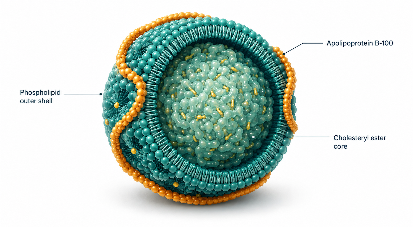
LDL particle physiology: a single apoB-100 molecule wraps each particle, which is why particle count and cholesterol content are not the same measurement.
A patient sits across from me holding a lab report with one number circled in red ink. LDL cholesterol: 168 mg/dL. Nobody has explained to him what LDL is, what it carries, where it comes from, or what determines whether it causes harm. He has been told only that the number is high and that a medication will lower it. He has been managing a laboratory value for four years without ever being told what the value measures.
This is the strangest feature of modern cholesterol care. The single most-discussed marker in preventive medicine is also the one patients understand least, and the confusion is not accidental. LDL cholesterol is not a substance. It is not a type of particle. It is an estimate of how much cholesterol is being carried inside a class of lipoproteins, and it tells you almost nothing about how many of those particles exist, what condition they are in, or what metabolic environment they are circulating through. Understanding LDL particle physiology — the actual biology of the particle rather than the arithmetic of the number — changes what you look for on a lab panel and what you decide to do about it.
What You Will Learn
- What an LDL particle is structurally, and what it transports besides cholesterol
- How LDL is produced from VLDL rather than secreted directly by the liver
- How the LDL receptor clears particles from circulation and what regulates it
- Why insulin resistance changes LDL particle size, number, and residence time
- What determines whether a circulating LDL particle enters and stays in the arterial wall
- Why LDL cholesterol and LDL particle number frequently disagree, and which one tracks risk
What an LDL Particle Actually Is
An LDL particle is a transport vehicle, not a molecule of cholesterol. It is a roughly spherical structure about 20 to 25 nanometres across, with an outer shell of phospholipids, free cholesterol, and a single large structural protein called apolipoprotein B-100. Inside that shell sits a hydrophobic core of cholesteryl esters and triglycerides. Because lipids are not water-soluble, they cannot travel through plasma unaccompanied. The lipoprotein exists to solve that problem: it wraps a water-insoluble cargo in a water-compatible surface so it can move through the bloodstream to tissues that need it.
The apoB-100 protein is the defining feature. Each LDL particle carries exactly one apoB molecule, and that molecule is never exchanged or transferred between particles. This one-to-one relationship is the reason apoB measurement gives a direct count of atherogenic particles, a point I develop in detail on the cornerstone page for this cluster, cholesterol and cardiovascular risk. ApoB also functions as the address label: it is the ligand that allows the particle to be recognised and taken up by cells.
What LDL carries matters as much as the fact that it carries anything. Cholesterol is a structural requirement of every cell membrane in the body, the precursor for cortisol, aldosterone, testosterone, oestradiol, and vitamin D, and a component of bile acids and myelin. LDL also transports fat-soluble vitamins and carotenoids, and contributes to innate immune function by binding and neutralising bacterial endotoxin.
A physiology that evolved to deliver these things to every tissue in the body is not, in itself, pathological. The particle is doing its job. What varies between people is how many particles are in circulation, how long they stay there, what condition they are in, and whether the arterial wall they pass through is receptive to retaining them.
This is where the framing usually goes wrong in clinical practice. The question “is your LDL high” treats a transport system as a toxin. The more useful question is why the transport system is operating at the volume and under the conditions that it is — which is almost always a question about the metabolic state of the liver, and therefore a question about insulin resistance.
Where LDL Comes From: The VLDL Cascade
The liver does not secrete LDL. It secretes very-low-density lipoprotein, and LDL is what remains after VLDL has been progressively stripped of its triglyceride cargo in circulation.
A VLDL particle leaves the liver carrying one apoB-100 molecule and a large triglyceride-rich core. Its job is to export fat from the liver to peripheral tissue. As it travels, lipoprotein lipase on the capillary endothelium of muscle and adipose tissue hydrolyses the triglycerides inside it, releasing fatty acids for oxidation or storage. As the triglyceride core empties, the particle shrinks and its density rises. It passes through an intermediate-density stage and, if delipidation continues, ends as an LDL particle: smaller, denser, and now cholesterol-enriched relative to its remaining triglyceride content.
Two things follow from this that are rarely made explicit to patients. First, the number of LDL particles in circulation is determined largely by the rate of hepatic VLDL secretion, not by dietary cholesterol intake. If the liver is exporting more apoB-containing particles, more LDL particles will exist downstream, regardless of how much cholesterol is on the plate. Second, LDL particle composition depends on what happened during the cascade — how much triglyceride the particle started with, how efficiently it was hydrolysed, and how much lipid exchange occurred along the way.
That exchange step is central. Cholesteryl ester transfer protein moves cholesteryl esters out of LDL and HDL and moves triglycerides in, in a swap that intensifies when plasma triglyceride concentrations are high.
An LDL particle that has been loaded with triglyceride through this exchange becomes a substrate for hepatic lipase, which hydrolyses that triglyceride and leaves behind a particle that is smaller, denser, and depleted of cholesterol relative to where it started. The upstream driver of this entire sequence is hepatic triglyceride availability — which is why LDL physiology cannot be understood separately from what is happening in the liver, and why the pathway that produces fatty liver from insulin resistance is also the pathway that reshapes the LDL profile.
The clinical consequence is that two people with identical LDL cholesterol can have arrived there by entirely different routes: one through a modest number of large, cholesterol-rich particles, the other through a much larger number of small, cholesterol-poor ones. The cholesterol assay cannot distinguish between them, because it measures the cargo and not the carriers.
How LDL Leaves the Circulation
LDL is cleared from plasma primarily by the LDL receptor, a cell-surface protein that was characterised in the work that earned Brown and Goldstein the 1985 Nobel Prize and that established receptor-mediated endocytosis as a general principle in cell biology.
The mechanism is elegant. LDL receptors cluster in clathrin-coated pits on the hepatocyte surface. When an LDL particle binds — via apoB-100 — the pit invaginates and pinches off into the cell as a coated vesicle. Inside the acidified endosome, the particle dissociates from the receptor. The receptor recycles back to the cell surface, while the particle is delivered to the lysosome, where cholesteryl esters are hydrolysed and free cholesterol is released into the cell.
That released cholesterol then regulates the system that produced it. When intracellular cholesterol is abundant, the SREBP-2 transcription factor stays bound in the endoplasmic reticulum membrane and LDL receptor synthesis falls. When intracellular cholesterol is scarce, SREBP-2 is cleaved and travels to the nucleus, and LDL receptor expression rises. This is a feedback loop: the cell adjusts its own uptake capacity according to its own supply. Statins work by exploiting it — by inhibiting cholesterol synthesis, they lower intracellular cholesterol, which upregulates LDL receptors, which increases clearance of LDL from plasma.
Receptor availability, therefore, is one of the two main determinants of plasma LDL concentration. The other is production rate. A person can have elevated LDL because their liver is exporting apoB particles at a high rate, because their receptor-mediated clearance is impaired, or because of both. Familial hypercholesterolaemia represents the pure clearance defect: mutations reducing LDL receptor function raise LDL cholesterol dramatically and, in a clear dose-dependent relationship, raise atherosclerotic risk. The genetic evidence in the other direction is equally consistent — variants that lower lifetime LDL exposure lower risk correspondingly.
Residence time matters as much as concentration. A particle that is cleared within a day and a half has fewer opportunities to interact with the arterial wall than one that circulates for four or five days. Small, dense LDL particles have reduced affinity for the LDL receptor and are cleared more slowly, which means they spend longer in circulation, are exposed to oxidative modification for longer, and have more chances to enter the subendothelial space. Two people can have the same particle production rate and very different arterial exposure because of this.
Why LDL Particle Physiology Changes Under Insulin Resistance
This is where the pathway becomes clinically decisive, and where the standard lipid panel becomes most misleading.
When hepatic insulin signalling is impaired, several things happen at once. De novo lipogenesis is upregulated, increasing hepatic triglyceride synthesis. Suppression of adipose lipolysis fails, so free fatty acid delivery to the liver rises. Apolipoprotein B degradation within the hepatocyte is reduced, so more apoB survives to be secreted. The result is increased secretion of large, triglyceride-rich VLDL particles.
Those particles feed the cascade described above. High plasma triglyceride drives CETP-mediated exchange, loading LDL and HDL with triglyceride and stripping them of cholesteryl ester. Hepatic lipase, whose activity is elevated in insulin-resistant states, then hydrolyses that triglyceride, leaving small, dense LDL particles behind and generating small HDL particles that are catabolised rapidly, which is why HDL cholesterol falls in the same patients.
The resulting phenotype is coherent and recognisable: raised triglycerides, low HDL cholesterol, and a predominance of small, dense LDL. Reaven’s group demonstrated the underlying relationship directly, showing that individuals with the small-dense LDL pattern had significantly greater insulin responses to oral glucose and measurably impaired insulin-mediated glucose disposal compared with those with the large-buoyant pattern. Austin’s case-control work had already established that the small-dense pattern carried roughly a threefold increase in myocardial infarction risk, independent of age, sex, and relative weight.
Note what LDL cholesterol does in this setting. Because small dense particles carry less cholesterol each, a person can double their particle number while their LDL cholesterol stays flat or even falls. The panel looks reassuring precisely when the particle burden is rising. This is the single most consequential blind spot in routine lipid screening, and it is systematic rather than occasional — it appears in exactly the population where cardiovascular risk is accumulating fastest.
The direction of causation also matters for treatment. Small dense LDL is not an independent disease requiring its own intervention; it is a downstream readout of hepatic triglyceride overproduction. Krauss’s controlled feeding work demonstrated this cleanly: moderate carbohydrate restriction improved the atherogenic dyslipidaemia — including LDL particle size and distribution — with effects comparable to those of weight loss and not dependent on it. Treat the hepatic lipid flux, and the LDL profile reorganises. Treat the LDL number in isolation, and the flux continues.

From Circulating Particle to Arterial Lesion
A circulating LDL particle causes no harm. Atherosclerosis begins when a particle crosses the endothelium, enters the subendothelial space, and is retained there.
Retention is a physical binding event. ApoB-100 contains basic amino acid sequences that bind ionically to the negatively charged sulphate groups on arterial wall proteoglycans. This is the response-to-retention model of early atherogenesis, and the experimental evidence for it is unusually direct: mice engineered to express LDL with mutated proteoglycan-binding sites developed substantially less atherosclerosis than mice expressing normal LDL, despite comparable lipoprotein levels. Binding affinity, not merely plasma concentration, determined whether disease developed.
Once retained, the particle is subject to modification. It undergoes oxidation, glycation, and enzymatic alteration, and it aggregates with other retained particles. Modified LDL is recognised by macrophage scavenger receptors, which — unlike the LDL receptor — are not downregulated by intracellular cholesterol accumulation. The macrophage therefore keeps ingesting modified particles until it becomes a lipid-laden foam cell. Foam cell formation, subsequent apoptosis, impaired clearance of dying cells, and accumulation of extracellular lipid and debris together build the necrotic core of the plaque.
Every step of this sequence is modulated by the metabolic environment. Small dense particles penetrate the endothelium more readily and bind proteoglycans with higher affinity. Glycation in the setting of chronic hyperglycaemia increases susceptibility to oxidative modification and impairs receptor-mediated clearance. Endothelial dysfunction — driven by hyperinsulinaemia, oxidative stress, and inflammation — increases permeability to lipoprotein entry in the first place. Chronic low-grade inflammation amplifies the macrophage response to whatever is retained.
So the same number of circulating particles produces meaningfully different arterial consequences depending on the biology surrounding them. This is not an argument that particle number is unimportant. It is an argument that particle number and metabolic environment are both necessary parts of the picture, and that clinical practice has largely operationalised only one of them — and has operationalised it through a proxy measure that does not reliably track it.
Why LDL Cholesterol and LDL Particle Number Disagree
LDL cholesterol is not measured directly in most laboratories. It is calculated, and what it estimates is the mass of cholesterol contained within the LDL fraction. LDL particle number, whether measured as apoB or by NMR, counts particles.
If every LDL particle contained the same amount of cholesterol, the two measures would be interchangeable. They do not. Cholesterol content per particle varies substantially, and it varies most in exactly the patients whose risk is hardest to assess. When particles are cholesterol-depleted, particle number runs high relative to LDL cholesterol. This is discordance, and it is common.
Discordance has been characterised in large cohorts. In the Framingham Offspring Study, participants whose LDL particle number was high while their LDL cholesterol was at target had substantially higher cardiovascular event rates than participants whose particle number was low — the risk tracked the particle count rather than the cholesterol mass. A meta-analysis spanning more than 233,000 subjects and nearly 23,000 events found the same ranking: apoB was the strongest of the three markers, non-HDL cholesterol intermediate, and LDL cholesterol the weakest.
Practically, this means that when the metabolic context suggests discordance is likely, LDL cholesterol alone is not sufficient. The triglyceride-to-HDL ratio is a useful screening proxy: a ratio at or above roughly 3.8 has been shown to identify the small-dense LDL phenotype with high specificity, which makes it a reasonable trigger for ordering apoB. Fasting insulin, HOMA-IR, hepatic markers, and hs-CRP fill in the rest of the picture. None of this replaces LDL as a consideration. It contextualises it — which is what a transport system requires and what a single calculated number cannot supply.
Clinical Perspective: What I See in Practice
One of the most common patterns I see is a patient referred because of an elevated LDL-C, often already taking a statin, while the underlying metabolic picture has received very little attention.
When I look beyond LDL-C, I frequently find elevated fasting insulin, high triglycerides, low HDL cholesterol, central adiposity, and evidence of hepatic insulin resistance. In other words, the patient has the classic phenotype of metabolic syndrome. At that point, LDL-C is no longer the first question I ask. Instead, I want to know how many ApoB-containing particles are circulating, whether the LDL particles are predominantly small and dense, and whether the patient has the metabolic environment that promotes lipoprotein retention, oxidation, and endothelial dysfunction. LDL particles are simply transport vehicles. They become clinically relevant when they exist in a physiological context that favours arterial injury.
Consider two patients with an identical LDL-C of 180 mg/dL. The first has low fasting insulin, triglycerides of 70 mg/dL, HDL of 70 mg/dL, normal ApoB, low hs-CRP, good glycaemic control, and no features of insulin resistance. The second has fasting insulin of 20 µIU/mL, triglycerides of 300 mg/dL, HDL of 35 mg/dL, elevated ApoB, fatty liver, and chronic hyperglycaemia. The LDL-C number is identical. The physiology is not.
What I tell patients is this. LDL is not inherently a harmful substance. It is an essential lipoprotein that transports cholesterol, fat-soluble vitamins, and other lipids to tissues throughout the body. The real question is not simply how much cholesterol is inside LDL particles, but what metabolic environment those particles are circulating in. When insulin resistance develops, the liver overproduces VLDL, triglyceride exchange between lipoproteins increases, LDL particles often become smaller and denser, and the arterial wall becomes more susceptible to lipoprotein retention and inflammation. In that setting, cardiovascular risk is driven by the interaction between lipoproteins and metabolic dysfunction, not by LDL-C in isolation.
So rather than treating a cholesterol number alone, my goal is to understand why that lipoprotein profile exists. Improving insulin sensitivity, reducing hepatic fat accumulation, lowering ApoB when appropriate, and restoring metabolic health often changes the entire lipid pattern — not just the LDL-C value. That is a far more informative and physiologically meaningful approach than interpreting LDL-C in isolation.
Clinical Implications
If LDL is a transport system rather than a toxin, several things follow for how a lipid panel should be read and what should be done about it.
The first implication concerns what gets measured. An LDL cholesterol value interpreted without triglycerides, HDL cholesterol, fasting insulin, and hepatic markers alongside it is an incomplete measurement, because the same LDL-C carries a different meaning depending on the metabolic state producing it. Where the triglyceride-to-HDL ratio sits at or above roughly 3.8, where central adiposity is present, or where hepatic enzymes are drifting upward, discordance between cholesterol mass and particle number becomes likely enough that apoB should be measured rather than inferred. ApoB is inexpensive, standardized, and directly counts the particles that matter. Ordering it converts a probabilistic argument into a number.
The second implication concerns sequence. Because small dense LDL and elevated particle number are downstream consequences of hepatic triglyceride overproduction, intervening at the level of hepatic lipid flux addresses the whole pattern rather than one component of it. In practice this means addressing insulin resistance first: carbohydrate load, hepatic fat, protein adequacy, muscle mass, sleep, and circadian regularity. When hepatic VLDL secretion falls, triglycerides fall, HDL rises, particle size increases, and particle number declines — a coherent reorganization rather than a single-marker change. Krauss’s work is the clearest demonstration that this reorganization does not require weight loss to occur.
The third implication concerns what lipid-lowering therapy does and does not accomplish. Lowering apoB particle concentration reduces atherosclerotic events, and the evidence for this is strong and consistent. But lowering the particle count does not by itself repair the endothelium, reduce hepatic fat, reverse glycation, or restore insulin sensitivity, and the arterial wall’s receptivity to retaining whatever particles remain is governed by those factors. A patient whose apoB has been halved while their fasting insulin remains at 20 µIU/mL has addressed one determinant of risk and left the other running. This is not an argument against lipid-lowering therapy. It is an argument that the two questions are separate and that answering one does not answer the other.
The fourth implication concerns monitoring. If the goal is metabolic restoration rather than a target number, the markers worth tracking over time are apoB, triglycerides, HDL, fasting insulin, HOMA-IR, and hepatic markers together — reviewed at six and twelve weeks and interpreted as a pattern. A falling LDL-C accompanied by unchanged fasting insulin and unchanged triglycerides means something quite different from the same fall accompanied by improvement in both.
A Note on Uncertainty
Several elements of this picture are well established and several are not. That LDL receptor function governs plasma LDL concentration, that apoB retention in the arterial intima initiates atherogenesis, and that lifetime apoB exposure relates causally and dose-dependently to atherosclerotic events are supported by convergent genetic, mechanistic, and interventional evidence.
Less settled is whether small dense LDL is independently more atherogenic than large buoyant LDL once particle number is fully accounted for. Because the small-dense phenotype travels with a higher particle count, multivariate analyses have often found the size effect attenuates when particle number is included. My clinical position is that the argument is less important than it appears: the phenotype reliably signals the hepatic and insulin biology that produced it, and that biology is what warrants intervention regardless of how the size-versus-number question resolves.
Also unsettled is how much residual risk persists after aggressive apoB lowering in patients whose insulin resistance remains untreated, and how to weight competing markers when they diverge in an individual patient. These are open questions. I would rather name them than present a cleaner picture than the evidence supports.

The Animal-Based Protocol for Insulin Resistance and Type 2 Diabetes
A Functional Medicine Framework
People Also Ask
What is the difference between LDL and LDL cholesterol?
LDL is a lipoprotein particle — a transport vehicle made of phospholipids, proteins, and lipid cargo. LDL cholesterol is the estimated mass of cholesterol carried inside all of those particles combined. The distinction matters because cholesterol content per particle is variable, so the same LDL cholesterol value can represent very different particle counts.
Is LDL actually bad cholesterol?
No. LDL is the delivery system that supplies cholesterol to every cell in the body, transports fat-soluble vitamins, and participates in innate immune defence. The relevant clinical questions are how many apoB-containing particles are circulating, how long they remain in circulation, what condition they are in, and whether the arterial wall is primed to retain them.
Why is my LDL high if I eat well?
Plasma LDL concentration is determined mainly by hepatic VLDL secretion and by LDL receptor–mediated clearance, not by dietary cholesterol. Insulin resistance raises VLDL output; genetic variation affects receptor function; thyroid status, weight change, and rapid fat loss all shift the balance. Diet quality matters, but it acts on these upstream mechanisms rather than directly on the number.
What is small dense LDL and why does it matter?
Small dense LDL particles are produced when high plasma triglyceride drives cholesteryl ester transfer out of LDL, followed by hepatic lipase hydrolysis of the triglyceride that replaced it. These particles bind arterial proteoglycans more readily, are cleared more slowly, and are more susceptible to oxidative modification. The pattern has been associated with roughly a threefold increase in myocardial infarction risk.
Should I ask for an ApoB test?
ApoB counts atherogenic particles directly, since each particle carries exactly one apoB-100 molecule. It is particularly informative when discordance is likely — elevated triglycerides, low HDL, central adiposity, elevated fasting insulin, or fatty liver. In those situations LDL cholesterol can substantially understate the circulating particle burden.
Can insulin resistance raise LDL particle number without raising LDL cholesterol?
Yes, and this is the most clinically consequential blind spot in routine screening. Because small dense particles carry less cholesterol each, particle number can rise while the calculated LDL cholesterol stays unchanged or falls. The lipid panel appears stable while arterial exposure increases.
What does the triglyceride to HDL ratio tell me about my LDL?
It functions as an accessible proxy for LDL particle size. A ratio at or above approximately 3.8 identifies the small-dense phenotype with high specificity, which makes it a practical trigger for ordering apoB or particle-number testing rather than relying on LDL cholesterol alone.
Does lowering LDL always reduce cardiovascular risk?
Genetic and trial evidence consistently shows that lowering apoB-containing particle concentration reduces atherosclerotic events in proportion to the absolute reduction and the duration of exposure. What remains contested is how much residual risk is driven by the metabolic environment — insulin resistance, hepatic fat, glycation, inflammation, endothelial dysfunction — that persists after the number has been lowered.
About the Author
Morteza Ariana is a State-Certified Functional Nutritionist based in Germany, specializing in insulin resistance, type 2 diabetes, and root-cause metabolic restoration. He holds advanced training in systems-based physiology and has worked with patients across the U.S. and Europe for over 10 years.
His clinical framework is built around a core principle that mainstream medicine consistently overlooks: chronically elevated insulin — not blood glucose — is the earliest and most actionable driver of metabolic disease. That conviction was shaped in part by his own experience with hyperinsulinemia in 2016, and deepened through a decade of clinical practice and the study of leading researchers in metabolic medicine including Benjamin Bikman, Joseph Kraft, Gerald Reaven, Jason Fung, and Stephen Phinney.
His work focuses on identifying and correcting the upstream metabolic signals — insulin load, liver-gut axis dysfunction, circadian misalignment, and micronutrient gaps — that standard screening misses entirely. Patient outcomes are documented, anonymized, and published on this site.
If this resonates, the next step is clarity
The Metabolic Restoration Blueprint is a structured 12-week framework designed to correct upstream metabolic drivers — not just manage symptoms.
References
Borén J, Chapman MJ, Krauss RM, et al. Low-density lipoproteins cause atherosclerotic cardiovascular disease: pathophysiological, genetic, and therapeutic insights: a consensus statement from the European Atherosclerosis Society Consensus Panel. Eur Heart J. 2020;41(24):2313-2330. PMID: 32052833
Brown MS, Goldstein JL. A receptor-mediated pathway for cholesterol homeostasis. Science. 1986;232(4746):34-47. PMID: 3513311
Goldstein JL, Brown MS, Anderson RG, Russell DW, Schneider WJ. Receptor-mediated endocytosis: concepts emerging from the LDL receptor system. Annu Rev Cell Biol. 1985;1:1-39. PMID: 2881559
Olofsson SO, Borén J. Apolipoprotein B: a clinically important apolipoprotein which assembles atherogenic lipoproteins and promotes the development of atherosclerosis. J Intern Med. 2005;258(5):395-410. PMID: 16238675
Williams KJ, Tabas I. The response-to-retention hypothesis of early atherogenesis. Arterioscler Thromb Vasc Biol. 1995;15(5):551-561. PMID: 7749869
Skålén K, Gustafsson M, Rydberg EK, Hultén LM, Wiklund O, Innerarity TL, Borén J. Subendothelial retention of atherogenic lipoproteins in early atherosclerosis. Nature. 2002;417(6890):750-754. PMID: 12066187
Tabas I, Williams KJ, Borén J. Subendothelial lipoprotein retention as the initiating process in atherosclerosis: update and therapeutic implications. Circulation. 2007;116(16):1832-1844. PMID: 17938300
Austin MA, Breslow JL, Hennekens CH, Buring JE, Willett WC, Krauss RM. Low-density lipoprotein subclass patterns and risk of myocardial infarction. JAMA. 1988;260(13):1917-1921. PMID: 3418853
Reaven GM, Chen YD, Jeppesen J, Maheux P, Krauss RM. Insulin resistance and hyperinsulinemia in individuals with small, dense low density lipoprotein particles. J Clin Invest. 1993;92(1):141-146. PMID: 8325978
Krauss RM, Blanche PJ, Rawlings RS, Fernstrom HS, Williams PT. Separate effects of reduced carbohydrate intake and weight loss on atherogenic dyslipidemia. Am J Clin Nutr. 2006;83(5):1025-1031. PMID: 16685042
Hanak V, Munoz J, Teague J, Stanley A Jr, Bittner V. Accuracy of the triglyceride to high-density lipoprotein cholesterol ratio for prediction of the low-density lipoprotein phenotype B. Am J Cardiol. 2004;94(2):219-222. PMID: 15246907
Cromwell WC, Otvos JD, Keyes MJ, Pencina MJ, Sullivan L, Vasan RS, Wilson PW, D’Agostino RB. LDL particle number and risk of future cardiovascular disease in the Framingham Offspring Study — implications for LDL management. J Clin Lipidol. 2007;1(6):583-592. PMID: 19657464
Sniderman AD, Williams K, Contois JH, Monroe HM, McQueen MJ, de Graaf J, Furberg CD. A meta-analysis of low-density lipoprotein cholesterol, non-high-density lipoprotein cholesterol, and apolipoprotein B as markers of cardiovascular risk. Circ Cardiovasc Qual Outcomes. 2011;4(3):337-345. PMID: 21487090
Ference BA, Ginsberg HN, Graham I, et al. Low-density lipoproteins cause atherosclerotic cardiovascular disease. 1. Evidence from genetic, epidemiologic, and clinical studies. A consensus statement from the European Atherosclerosis Society Consensus Panel. Eur Heart J. 2017;38(32):2459-2472. PMID: 28444290