
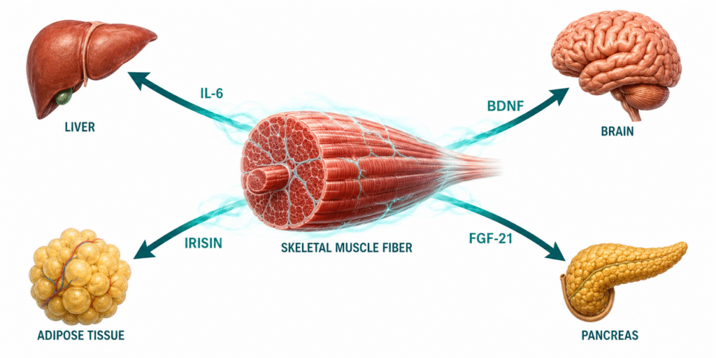
Myokines are the molecular signals contracting muscle sends to the liver, brain, pancreas, and fat tissue — and their absence is one of the most overlooked drivers of metabolic disease.
Your muscles have been sending messages your entire life. Every time a muscle fiber contracts — whether during a brisk walk, a resistance training session, or even sustained daily movement — it releases a class of signaling proteins into circulation that travel to distant organs and instruct them to behave differently. These proteins are called myokines, and they represent one of the most significant discoveries in metabolic medicine of the past two decades.
The concept is straightforward but its implications are not. Skeletal muscle is not a passive recipient of hormonal signals. It is an active producer of them. When it contracts regularly and under sufficient load, it broadcasts a coordinated molecular signal that
- reduces inflammation
- improves insulin sensitivity
- supports liver function
- protects pancreatic beta cells
- promotes fat oxidation
- supports brain health
When it does not contract — when it atrophies from disuse, aging, or chronic underloading — that signal goes quiet. What follows is not simply a loss of strength. It is the loss of a hormonal system that the rest of the body depends on.
This post maps the key myokines, their mechanisms, and their clinical relevance — and explains why the systemic improvements patients experience when they begin moving consistently are not coincidental.
What you will learn: What myokines are and how they are produced | The mechanisms and clinical significance of the most important individual myokines | Why myokine signaling explains improvements in mood, cognition, inflammation, and liver function that diet alone cannot fully account for | What this means practically for metabolic recovery
What Myokines Are and Why They Were Overlooked
For most of the twentieth century, endocrinology focused on glands — the pancreas, thyroid, adrenal cortex, pituitary — as the body’s hormonal infrastructure. Skeletal muscle, which comprises roughly 40 percent of total body mass in a healthy adult, was categorized as a target of hormonal signals rather than a source of them. That classification began to unravel in the early 2000s when Danish researcher Bente Klarlund Pedersen identified interleukin-6 being released from contracting muscle in quantities that could not be explained by immune activation alone. The muscle itself was the source.
That discovery opened a new field. The term myokine — from the Greek for muscle and motion — was coined to describe cytokines and peptides produced and secreted specifically by skeletal muscle in response to contraction. Since then, researchers have catalogued over six hundred candidate myokines. A well-characterized subset now has mapped mechanisms and documented clinical effects. The full picture continues to be assembled, but what is already known is sufficient to reframe what physical activity actually does at the molecular level.
What distinguishes myokines from classical hormones is their production logic. Traditional endocrine hormones are secreted by dedicated glandular tissue in response to defined biochemical stimuli. Myokines are secreted in proportion to mechanical load, contraction frequency, and exercise duration. They act locally within muscle tissue, regionally on adjacent adipose tissue, and systemically on the liver, pancreas, brain, bone, and immune system. Their output is determined entirely by how much the muscle is actually being used.
The Key Myokines: Mechanisms and Clinical Significance
Interleukin-6: Anti-Inflammatory Signal from Muscle, Pro-Inflammatory Signal from Fat
IL-6 is the most studied myokine and the most frequently misunderstood in clinical settings. On a standard inflammatory panel, elevated IL-6 is a marker of metabolic disease — and correctly so. But that is adipose-derived IL-6, produced by inflamed fat tissue, which drives the production of TNF-α and IL-1β and deepens systemic insulin resistance. It is a genuine inflammatory signal.
Muscle-derived IL-6, released in large quantities during sustained contraction, operates through a fundamentally different pathway. It stimulates fat oxidation in working muscle, enhances hepatic glucose production during exercise to maintain fuel availability, and — critically — suppresses TNF-α and IL-1β rather than stimulating them. It is anti-inflammatory. The same molecule carries opposing biological instructions depending entirely on whether it originates from contracting muscle or from dysfunctional adipose tissue.
This distinction has direct clinical relevance. In sedentary individuals or those with significant muscle loss, the anti-inflammatory IL-6 signal from muscle is absent. The pro-inflammatory IL-6 from adipose tissue is not. The net cytokine environment shifts toward chronic low-grade inflammation — one of the most consistent upstream drivers of insulin resistance, as detailed in the post on chronic low-grade inflammation and insulin resistance. Rebuilding the muscle-derived IL-6 signal is not a minor metabolic adjustment. It is a structural shift in the body’s inflammatory baseline.
Irisin: Fat Browning, Bone Protection, and Insulin Sensitivity
Irisin is cleaved from a membrane protein called FNDC5 and released predominantly during resistance exercise. Its primary documented action is the browning of white adipose tissue — the conversion of metabolically inert fat storage cells into beige adipocytes that dissipate energy as heat rather than storing it. This thermogenic shift reduces visceral fat mass and the inflammatory burden that accompanies it, which is itself a major upstream driver of hepatic insulin resistance.
Beyond adipose tissue, irisin has documented protective effects on bone mineral density — particularly relevant in older patients where muscle loss and osteoporosis frequently coexist — and appears to mediate some of the cognitive benefits of resistance training through its stimulation of BDNF expression in the hippocampus. Its production is substantially reduced in sedentary individuals and rises in proportion to mechanical loading during exercise.
BDNF: The Molecular Bridge Between Muscle and Brain
Brain-derived neurotrophic factor is produced in the central nervous system but is also secreted by contracting skeletal muscle and crosses the blood-brain barrier. During exercise, circulating BDNF rises substantially, and this increase mediates well-documented improvements in memory, executive function, and mood regulation. It also plays a role in hypothalamic regulation of energy balance and appetite — its reduction with inactivity and muscle loss may contribute to the dysregulated hunger signaling that complicates metabolic recovery.
The clinical significance here is that the mood stabilization, cognitive clarity, and emotional resilience that patients report when they begin moving consistently are not purely psychological responses to lifestyle change. They reflect a measurable neurochemical shift driven in part by muscle contraction. BDNF is one of the biological links that connects regular movement to mental function — a connection that nutrition alone, however well structured, cannot fully replicate.
FGF-21: Metabolic Flexibility at the Cellular Level
Fibroblast growth factor 21 is produced by multiple tissues, but skeletal muscle secretes it during both exercise and fasting states. Muscle-derived FGF-21 acts locally to enhance fatty acid oxidation and improve mitochondrial function — the cellular machinery that determines whether a cell can shift efficiently between glucose and fat as fuel sources. This is the molecular substrate of metabolic flexibility, the capacity to use the right fuel at the right time that deteriorates progressively in insulin-resistant individuals. The relationship between FGF-21, mitochondrial adaptation, and the ability to sustain fat oxidation at moderate exercise intensities is examined in detail in the post on metabolic flexibility.
IGF-1: Local Muscle Repair and Mass Maintenance
IGF-1 produced locally within muscle tissue in response to mechanical loading drives muscle protein synthesis and satellite cell activation — the cellular repair machinery that maintains and rebuilds muscle mass after training. Unlike hepatic IGF-1, which is systemically regulated, local muscle IGF-1 is responsive to the immediate mechanical environment. Underload the muscle chronically, and local IGF-1 production falls, impairing mass maintenance independent of systemic hormonal status. This is one reason why muscle loss in sedentary aging accelerates independently of testosterone or growth hormone decline — the local anabolic signal has been removed by disuse.
Myonectin and IL-15: Lipid Clearance and Lean Mass
Myonectin, secreted following exercise, promotes fatty acid uptake in the liver and adipose tissue — contributing to the coordinated post-exercise clearance of circulating triglycerides. IL-15, another contraction-induced myokine, promotes skeletal muscle hypertrophy and has documented anti-adipogenic effects, suppressing fat cell development and supporting lean mass accrual. Both myokines represent components of the post-exercise metabolic window that is frequently underutilized in patients who train without adequate protein intake or who skip the recovery period entirely.

Clinical Perspective: What I See in Practice
What I observe consistently in clinical practice is that the improvements patients experience when they begin moving regularly — particularly through Zone 2 walking and structured resistance training — extend well beyond what blood sugar or body weight numbers capture.
Patients describe better mood, clearer thinking, more stable energy across the day, improved sleep, and faster recovery from physical exertion. These changes frequently appear before significant weight loss has occurred. It is tempting to attribute them entirely to nutrition improvements or to the psychological effect of taking action. I do not think that tells the whole story.
When I look at the inflammatory markers alongside metabolic ones, a pattern emerges. As insulin sensitivity improves and consistent movement becomes established, hsCRP declines — often meaningfully — alongside the expected improvements in ALT, GGT, and the TG/HDL ratio. The inflammatory signal is moving in a direction that diet alone, in my clinical experience, does not reliably produce at the same pace. The addition of regular muscle contraction appears to be doing something distinct.
I am careful not to attribute all of this specifically to myokines. In real clinical practice, patients improve multiple variables simultaneously: nutrition, sleep, stress load, body composition, and movement. These factors interact. What I can say is that the myokine framework provides a coherent biological explanation for why the benefits of regular movement are so consistently systemic — why a patient who begins walking daily and lifting twice a week reports sleeping better, thinking more clearly, and recovering faster from illness, even when the scale has barely moved. Muscle contraction is not producing one localized effect. It is rewriting the hormonal environment the entire body operates in.
The patients in whom I see the slowest myokine-related recovery are those who exercise intensely but remain sedentary for the other twenty-two hours of the day, those whose training is dominated by high-intensity cardio without sufficient resistance stimulus, and those whose nutritional environment keeps insulin elevated around the clock, suppressing the fat oxidation and inflammatory resolution that myokines are attempting to facilitate. The signal is being produced but cannot be received.
Why Myokine Output Declines — and What That Costs
Myokine production is not a fixed biological constant. It is a use-dependent output. Three conditions reliably reduce it: muscle loss from aging or chronic disuse, insufficient contraction frequency across the day, and training structures that do not generate adequate mechanical tension.
Sarcopenia — the progressive loss of muscle mass from the fourth decade onward — reduces myokine output not just because there is less muscle tissue to contract, but because the remaining tissue contracts less forcefully and less frequently. The anti-inflammatory IL-6, the irisin-driven fat browning, the FGF-21-mediated mitochondrial signaling, the BDNF-supported neurological function — all of it declines proportionally. As covered in the post on skeletal muscle and metabolic health, sarcopenia is not a geriatric inconvenience. It is a metabolic disease with systemic hormonal consequences.
The liver bears a disproportionate share of that cost. Muscle-derived IL-6 and FGF-21 both modulate hepatic lipid metabolism and glucose output. When those signals disappear, hepatic insulin resistance deepens and de novo lipogenesis accelerates. The pancreas loses the trophic support from irisin and IL-6 that helps maintain beta cell function. The brain loses the BDNF stimulus that sustains neuroplasticity and cognitive reserve. These are not theoretical downstream risks. They are the biological substrate of the metabolic deterioration that accumulates silently over years of inactivity.

The Animal-Based Protocol for Insulin Resistance and Type 2 Diabetes
A Functional Medicine Framework
A Note on Uncertainty
The myokine field remains active and incompletely mapped. While IL-6, irisin, BDNF, FGF-21, IGF-1, and myonectin have well-supported mechanisms in human physiology, many of the over six hundred candidate myokines identified to date have not had their mechanisms or clinical significance fully characterized. Some findings from animal models have not translated directly to human outcomes. The precise dose-response relationships between specific exercise modalities, training volumes, and myokine output profiles are still being established. The directional evidence is robust: regular muscle contraction produces systemic anti-inflammatory, insulin-sensitizing, and organ-protective signals, and the loss of that signaling has measurable metabolic consequences. The full mechanistic map continues to be drawn.
People Also Ask
What are myokines?
Myokines are cytokines and peptides produced and secreted by skeletal muscle in response to contraction. They act locally within muscle tissue and systemically on distant organs including the liver, brain, pancreas, adipose tissue, and bone, regulating inflammation, insulin sensitivity, fat metabolism, and neurological function.
How do myokines affect insulin resistance?
Myokines including IL-6, irisin, and FGF-21 improve insulin sensitivity through multiple mechanisms: reducing inflammatory cytokines that impair insulin signaling, promoting fat oxidation in muscle and adipose tissue, supporting hepatic lipid metabolism, and enhancing mitochondrial function. Their absence with muscle loss or inactivity removes a significant biological counter to insulin resistance progression.
Is IL-6 inflammatory or anti-inflammatory?
Both, depending on its source. IL-6 produced by inflamed adipose tissue drives TNF-α and IL-1β production and is pro-inflammatory. IL-6 produced by contracting skeletal muscle suppresses TNF-α and IL-1β and acts as an anti-inflammatory metabolic signal. The source determines the biological effect entirely.
What does irisin do in the body?
Irisin promotes the browning of white adipose tissue, improving fat oxidation and reducing visceral fat mass. It also supports bone mineral density, stimulates BDNF expression in the brain, and improves insulin sensitivity in peripheral tissues. Its production rises with resistance exercise and falls with inactivity.
Why do people feel mentally better when they exercise regularly?
Part of the explanation is myokine-mediated. Contracting muscle releases BDNF and irisin, both of which cross the blood-brain barrier and support neuroplasticity, mood regulation, and cognitive function. This neurological benefit is distinct from the effects of weight loss or improved glucose control and occurs through direct muscle-to-brain signaling.
Which exercise produces the most myokines?
Resistance training under sufficient mechanical load produces the strongest irisin and IGF-1 response. Sustained moderate-intensity activity generates the largest IL-6 response. Both modalities produce BDNF and FGF-21. Consistency and total daily contraction time across the week determine cumulative myokine output more than any single session’s intensity.
Can myokine production be impaired even in people who exercise?
Yes. Chronically elevated insulin from frequent carbohydrate intake, poor sleep, high inflammatory load, and excessive high-intensity training without adequate recovery can all reduce the effectiveness of myokine signaling — not by preventing secretion but by creating a metabolic environment in which the signals cannot be fully received.
About the Author
Morteza Ariana is a State-Certified Functional Nutritionist based in Germany, specializing in insulin resistance, type 2 diabetes, and root-cause metabolic restoration. He holds advanced training in systems-based physiology and has worked with patients across the U.S. and Europe for over 10 years.
His clinical framework is built around a core principle that mainstream medicine consistently overlooks: chronically elevated insulin — not blood glucose — is the earliest and most actionable driver of metabolic disease. That conviction was shaped in part by his own experience with hyperinsulinemia in 2016, and deepened through a decade of clinical practice and the study of leading researchers in metabolic medicine including Benjamin Bikman, Joseph Kraft, Gerald Reaven, Jason Fung, and Stephen Phinney.
His work focuses on identifying and correcting the upstream metabolic signals — insulin load, liver-gut axis dysfunction, circadian misalignment, and micronutrient gaps — that standard screening misses entirely. Patient outcomes are documented, anonymized, and published on this site.
If this resonates, the next step is clarity
The Metabolic Restoration Blueprint is a structured 12-week framework designed to correct upstream metabolic drivers — not just manage symptoms.
Scientific References
Raschke S, Eckel J. Adipo-myokines: two sides of the same coin — mediators of inflammation and mediators of exercise. Mediators Inflamm. 2013;2013:320724. 🔗 https://pubmed.ncbi.nlm.nih.gov/23737657/
Pedersen BK, Febbraio MA. Muscles, exercise and obesity: skeletal muscle as a secretory organ. Nat Rev Endocrinol. 2012;8(8):457–465. 🔗 https://pubmed.ncbi.nlm.nih.gov/22473333/
Pedersen BK. Muscle as a secretory organ. Compr Physiol. 2013;3(3):1337–1362. 🔗 https://pubmed.ncbi.nlm.nih.gov/23897689/
Severinsen MCK, Pedersen BK. Muscle-organ crosstalk: the emerging roles of myokines. Endocr Rev. 2020;41(4):594–609. 🔗 https://pubmed.ncbi.nlm.nih.gov/32393961/
Boström P, Wu J, Jedrychowski MP, et al. A PGC1-α-dependent myokine that drives brown-fat-like development of white fat and thermogenesis. Nature. 2012;481(7382):463–468. 🔗 https://pubmed.ncbi.nlm.nih.gov/22237023/
Wrann CD, White JP, Salogiannnis J, et al. Exercise induces hippocampal BDNF through a PGC-1α/FNDC5 pathway. Cell Metab. 2013;18(5):649–659. 🔗 https://pubmed.ncbi.nlm.nih.gov/24120943/
Pedersen BK. Physical activity and muscle-brain crosstalk. Nat Rev Neurosci. 2019;20(7):419–430. 🔗 https://pubmed.ncbi.nlm.nih.gov/31048807/
Febbraio MA, Pedersen BK. Contraction-induced myokine production and release: is skeletal muscle an endocrine organ? Exerc Sport Sci Rev. 2005;33(3):114–119. 🔗 https://pubmed.ncbi.nlm.nih.gov/16006818/
Schnyder S, Handschin C. Skeletal muscle as an endocrine organ: PGC-1α, myokines and exercise. Bone. 2015;80:115–125. 🔗 https://pubmed.ncbi.nlm.nih.gov/26453501/
Kim HJ, So B, Choi M, et al. Resistance exercise training increases the expression of irisin concomitant with improvement of muscle function in aging mice and humans. Exp Gerontol. 2015;70:11–17. 🔗 https://pubmed.ncbi.nlm.nih.gov/26169350/
Catoire M, Kersten S. The search for exercise factors in humans. FASEB J. 2015;29(5):1615–1628. 🔗 https://pubmed.ncbi.nlm.nih.gov/25609425/
Brandt C, Pedersen BK. The role of exercise-induced myokines in muscle homeostasis and the defense against chronic diseases. J Biomed Biotechnol. 2010;2010:520258. 🔗 https://pubmed.ncbi.nlm.nih.gov/20224659/
Ahima RS, Park HK. Connecting myokines and metabolism. Endocrinol Metab (Seoul). 2015;30(3):235–245. 🔗 https://pubmed.ncbi.nlm.nih.gov/26354480/
Nielsen AR, Pedersen BK. The biological roles of exercise-induced cytokines: IL-6, IL-8, and IL-15. Appl Physiol Nutr Metab. 2007;32(5):833–839. 🔗 https://pubmed.ncbi.nlm.nih.gov/18059593/