
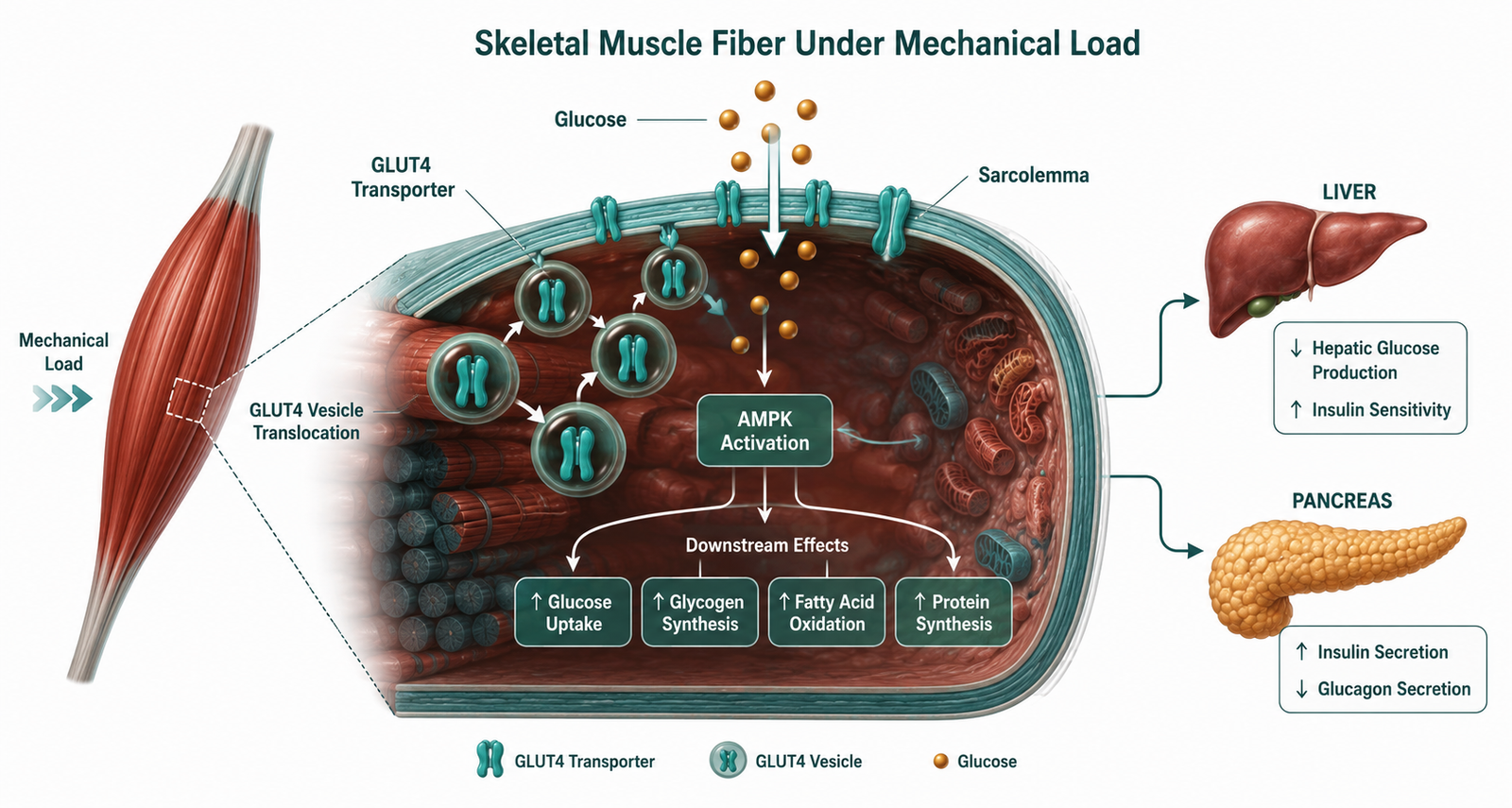
Resistance training and metabolism converge at the muscle cell: contraction drives GLUT4 to the surface independently of insulin, restoring glucose disposal from the first session.
Resistance training is discussed in mainstream medicine primarily as a tool for building strength, preventing falls in older adults, or improving body composition. What that framing misses is the metabolic dimension — and the metabolic dimension is where the real clinical story lives.
When a patient with insulin resistance begins a structured resistance training program, something predictable happens. Glucose disposal improves within hours of the first session. Fasting insulin begins to decline within weeks. Triglycerides follow. Liver enzymes normalize over months. Waist circumference shrinks before the scale moves significantly. And patients consistently report better energy, clearer thinking, fewer sugar cravings, and faster recovery from physical activity — often before a single laboratory value has changed on paper.
None of this is coincidental. Skeletal muscle is the body’s largest glucose disposal organ, and resistance training is the most direct intervention available for restoring its function. Understanding why requires looking at what happens at the cellular level when muscle contracts under load — and what is lost when it does not.
What you will learn: How resistance training restores insulin sensitivity through both acute and chronic mechanisms | Which metabolic markers respond first, and on what timeline | Why strength itself is a metabolic biomarker | How resistance training interacts with the liver, pancreas, and inflammatory system | What a practical resistance training protocol looks like for a metabolically compromised patient
How Resistance Training and Metabolism Are Linked at the Cellular Level
The primary mechanism is GLUT4 translocation. Under resting conditions, the glucose transporter protein GLUT4 sits in intracellular storage vesicles within muscle cells, waiting for an insulin signal to drive it to the cell surface. When insulin binds its receptor, a signaling cascade unfolds that moves GLUT4 transporters to the membrane, where they pull glucose out of circulation and into the cell.
Insulin resistance means that cascade is impaired. The signal is sent but poorly received. The pancreas compensates by producing more insulin — the compensatory hyperinsulinemia that precedes overt glucose dysregulation by a decade or more, as described in detail in the insulin resistance cornerstone.
Resistance training bypasses that impaired signaling entirely. Muscle contraction drives GLUT4 translocation through a parallel, insulin-independent pathway mediated by AMP-activated protein kinase (AMPK) and calcium signaling. The muscle pulls glucose out of circulation without waiting for insulin’s instruction. This is why a single resistance training session can meaningfully improve postprandial glucose disposal for the following 24 to 48 hours — the acute effect is real, immediate, and clinically significant even before any structural adaptation has occurred.
The chronic adaptations that follow sustained training are equally important. Regular resistance training increases total GLUT4 protein content in muscle tissue, expands the cross-sectional area of individual muscle fibers, and improves the sensitivity of the insulin signaling cascade itself. Over weeks and months, the muscle becomes a more efficient glucose sink under both exercise and resting conditions. The pancreas no longer needs to produce as much insulin to maintain fasting glucose. Compensatory hyperinsulinemia begins to resolve.
What Changes First: A Clinical Marker Timeline
One of the most instructive aspects of working with insulin-resistant patients through a resistance training protocol is learning which markers move first — because the sequence tells a story about the underlying mechanisms.
Postprandial glucose is frequently the earliest objective signal. Because contracting muscle acts as a direct glucose sink through insulin-independent GLUT4 translocation, smaller glucose excursions after meals and faster return to baseline can appear after the very first session. Patients using continuous glucose monitors often notice this immediately. This acute effect lasts roughly 24 to 48 hours, which is one of the strongest arguments for training frequency — the metabolic window needs to be kept open consistently.
Fasting insulin typically begins to decline within two to six weeks when resistance training is combined with nutritional intervention. The mechanism is the progressive restoration of muscle insulin sensitivity described above: as peripheral glucose disposal improves, the pancreatic demand for compensatory insulin output decreases. By eight to twelve weeks, the decline is usually measurable on a standard panel. Since fasting insulin is the earliest and most sensitive upstream marker of metabolic dysfunction — more so than fasting glucose or HbA1c, which lag significantly — this is often the most clinically meaningful early signal.
Triglycerides and the TG/HDL ratio typically begin falling within four to twelve weeks, particularly when resistance training is combined with reduced refined carbohydrate intake, adequate protein, and improved sleep. HDL moves more slowly — often over several months — but the TG/HDL ratio as a composite marker of insulin sensitivity trends in the right direction reliably and earlier than most clinicians expect. Its role as a proxy for insulin resistance is examined in the post on the TG/HDL ratio.
Liver enzymes — ALT, AST, and GGT — typically lag behind improvements in insulin action. In patients with metabolic dysfunction-associated steatotic liver disease, meaningful changes in liver enzymes generally require eight to twenty-four weeks, depending on the degree of liver fat, visceral fat reduction, alcohol intake, and the extent of dietary intervention running alongside training. Resistance training alone produces improvement, but the combination with nutritional change generates a substantially larger effect. The mechanisms connecting muscle glucose disposal to hepatic fat accumulation are detailed in the post on insulin resistance and fatty liver.
Waist circumference is one of the most practically useful and most overlooked clinical markers in this context. Many patients lose visceral fat and reduce waist circumference before the scale changes meaningfully — reflecting preferential loss of the metabolically active intra-abdominal fat that drives hepatic insulin resistance. A reduction of even a few centimeters is clinically significant and often appears within six to twelve weeks.
HbA1c moves last. Because it reflects average glucose exposure over the lifespan of red blood cells — approximately three months — it is structurally slow to respond. Patients who have made genuine metabolic progress will often show improved fasting insulin, triglycerides, TG/HDL ratio, and waist circumference well before HbA1c reflects the change. Treating HbA1c as the primary outcome metric in the early months of a resistance training intervention leads to systematic underestimation of clinical progress.

Clinical Perspective: What I See in Practice
The marker sequence I just described maps closely to what I observe in practice, but the laboratory picture is only part of the story.
What patients report before their numbers move is often the first signal that the intervention is working: less afternoon fatigue, fewer sugar cravings, better sleep, improved mental clarity, and faster recovery from exercise. These are not soft outcomes. They reflect improving metabolic flexibility — the capacity to shift between glucose and fat as fuel sources that deteriorates with insulin resistance and begins to restore with consistent muscle loading.
The pattern I find most clinically instructive is when fasting insulin, triglycerides, TG/HDL ratio, and waist circumference all move together in the right direction, even while fasting glucose and HbA1c are still unremarkable. That constellation tells me the upstream metabolic machinery is responding. The downstream markers will follow.
One observation I return to repeatedly: patients who add resistance training to a well-structured nutritional protocol progress substantially faster than those doing nutrition alone. The combination is not additive — it appears to be synergistic. Diet removes the substrate excess driving hyperinsulinemia. Resistance training restores the peripheral disposal capacity that hyperinsulinemia had overwhelmed. Both levers need to move.
The patients who respond most slowly are those who train hard two or three times per week but remain largely sedentary for the rest of the day. Total daily muscle contraction time matters, not just peak session intensity. A patient who trains for forty-five minutes and then sits for eight hours is producing a fraction of the cumulative metabolic benefit of someone who trains consistently and maintains meaningful daily movement outside of formal sessions. The relationship between contraction frequency, myokine output, and systemic metabolic signaling is examined in the post on myokines and the broader endocrine function of muscle in the post on muscle as an endocrine organ.
Strength as a Metabolic Biomarker
One of the most underappreciated implications of the resistance training and metabolism research is that muscular strength itself functions as a metabolic biomarker. Multiple large prospective studies have linked greater grip strength and lower-body strength to lower risk of developing type 2 diabetes, independent of body weight and cardiovascular fitness. The relationship is not purely correlational — it reflects the fact that stronger muscle is generally more insulin-sensitive muscle, with higher GLUT4 content, greater mitochondrial density, and superior glucose disposal capacity.
In practice, this means that tracking strength progression — the weight lifted, the repetitions completed, the recovery between sets — gives clinically relevant information about metabolic trajectory. A patient who is getting steadily stronger over a twelve-week intervention is almost certainly improving their insulin sensitivity in parallel, even if their fasting glucose has not yet moved. Strength is not a proxy for metabolic health in every context, but within a structured metabolic intervention, consistent strength gains are a reliable signal that the underlying machinery is becoming more resilient.
The Practical Protocol: What Resistance Training for Metabolic Recovery Actually Looks Like
The evidence base for resistance training in insulin resistance points consistently toward a few structural principles.
Frequency of two to three sessions per week produces meaningful metabolic adaptation without accumulating the cortisol load that excessive training frequency can generate in already-stressed metabolic systems. Each session should target the major compound movement patterns — lower body pushing and pulling, upper body pushing and pulling, and hip hinge movements — that engage the largest muscle groups and generate the greatest total glucose disposal demand.
Intensity matters metabolically. Training at 65 to 80 percent of one-repetition maximum consistently outperforms low-load high-repetition training for improving insulin sensitivity and GLUT4 expression in the research literature. This does not mean every set needs to be taken to failure — but it does mean that the load needs to be sufficient to generate meaningful mechanical tension. Resistance band circuits and light bodyweight work produce some benefit, but they are not equivalent to loaded progressive resistance training for the metabolic outcomes described in this post.
Progressive overload — the systematic increase in load, volume, or difficulty over time — is the stimulus that drives continued adaptation. Without it, the metabolic benefit plateaus. This is one reason a structured program with tracked progression produces superior outcomes to informal gym attendance.
Protein intake at 1.6 grams per kilogram of ideal body weight supports the muscle protein synthesis that makes structural adaptation possible. Without adequate protein, training stimulus is present but the raw material for rebuilding is not. This is one of the most common gaps in patients who exercise regularly but remain metabolically stalled.

The Animal-Based Protocol for Insulin Resistance and Type 2 Diabetes
A Functional Medicine Framework
A Note on Uncertainty
The evidence base for resistance training in insulin resistance is robust, but individual response varies considerably. Factors including baseline muscle mass, degree of insulin resistance, sleep quality, hormonal status, and the quality of nutritional support all influence the rate and magnitude of metabolic adaptation. The timelines described in this post reflect clinical patterns rather than guaranteed individual outcomes. Some patients respond faster, some slower, and the combination of variables in any individual case makes precise prediction impossible. What the evidence consistently supports is the direction: resistance training improves insulin sensitivity, reduces compensatory hyperinsulinemia, and produces systemic metabolic benefits that no pharmaceutical intervention currently replicates with the same breadth of effect.
People Also Ask
Does resistance training improve insulin resistance?
Yes. Resistance training improves insulin resistance through two mechanisms: an acute insulin-independent increase in glucose disposal via GLUT4 translocation during and immediately after exercise, and chronic adaptations including increased GLUT4 protein content, greater muscle mass, and improved insulin signaling sensitivity that accumulate over weeks and months of consistent training.
How quickly does resistance training lower insulin levels?
Fasting insulin typically begins to decline within two to six weeks when resistance training is combined with nutritional intervention, with clearer improvements measurable by eight to twelve weeks. Postprandial glucose improvements can appear after the first session due to the acute GLUT4 effect.
Which is better for insulin resistance — cardio or resistance training?
Both have documented benefits, but resistance training produces a qualitatively different and more durable metabolic adaptation through increases in muscle mass and GLUT4 content. The combination of resistance training with moderate daily movement — including Zone 2 walking — produces superior outcomes to either modality alone.
What does resistance training do to the liver?
Resistance training improves hepatic insulin sensitivity by reducing the glucose and fatty acid substrate that would otherwise drive hepatic de novo lipogenesis. Combined with nutritional intervention, it reduces liver fat and normalizes ALT, AST, and GGT over eight to twenty-four weeks in patients with metabolic dysfunction-associated steatotic liver disease.
How many times per week should an insulin-resistant patient resistance train?
Two to three sessions per week targeting major compound movements at 65 to 80 percent of one-repetition maximum is the evidence-supported range for metabolic adaptation. Frequency beyond this can generate a counterproductive cortisol load in already-stressed metabolic systems without proportional additional benefit.
Is strength a sign of metabolic health?
In the context of a structured metabolic intervention, consistent strength gains reliably indicate improving insulin sensitivity. Stronger muscle generally has higher GLUT4 content, greater mitochondrial density, and superior glucose disposal capacity. Grip strength and lower-body strength are independently associated with reduced type 2 diabetes risk in large prospective studies.
Why do patients feel better before their lab results improve?
Because the earliest metabolic effects of resistance training — improved postprandial glucose disposal, reduced glucose excursions, better energy partitioning — occur at the cellular level before they register on standard fasting panels. Patients experience the functional improvement in energy, cognitive clarity, and recovery capacity before HbA1c or fasting glucose reflect the change.
About the Author
Morteza Ariana is a State-Certified Functional Nutritionist based in Germany, specializing in insulin resistance, type 2 diabetes, and root-cause metabolic restoration. He holds advanced training in systems-based physiology and has worked with patients across the U.S. and Europe for over 10 years.
His clinical framework is built around a core principle that mainstream medicine consistently overlooks: chronically elevated insulin — not blood glucose — is the earliest and most actionable driver of metabolic disease. That conviction was shaped in part by his own experience with hyperinsulinemia in 2016, and deepened through a decade of clinical practice and the study of leading researchers in metabolic medicine including Benjamin Bikman, Joseph Kraft, Gerald Reaven, Jason Fung, and Stephen Phinney.
His work focuses on identifying and correcting the upstream metabolic signals — insulin load, liver-gut axis dysfunction, circadian misalignment, and micronutrient gaps — that standard screening misses entirely. Patient outcomes are documented, anonymized, and published on this site.
If this resonates, the next step is clarity
The Metabolic Restoration Blueprint is a structured 12-week framework designed to correct upstream metabolic drivers — not just manage symptoms.
Scientific References
Srikanthan P, Karlamangla AS. Relative muscle mass is inversely associated with insulin resistance and prediabetes. J Clin Endocrinol Metab. 2011;96(9):2898–2903. 🔗 https://pubmed.ncbi.nlm.nih.gov/21697234/
Holten MK, Zacho M, Gaster M, et al. Strength training increases insulin-mediated glucose uptake, GLUT4 content, and insulin signaling in skeletal muscle in patients with type 2 diabetes. Diabetes. 2004;53(2):294–305. 🔗 https://pubmed.ncbi.nlm.nih.gov/14747278/
Ivy JL. Role of exercise training in the prevention and treatment of insulin resistance and non-insulin-dependent diabetes mellitus. Sports Med. 1997;24(5):321–336. 🔗 https://pubmed.ncbi.nlm.nih.gov/9368278/
Richter EA, Hargreaves M. Exercise, GLUT4, and skeletal muscle glucose uptake. Physiol Rev. 2013;93(3):993–1017. 🔗 https://pubmed.ncbi.nlm.nih.gov/23899560/
Strasser B, Siebert U, Schobersberger W. Resistance training in the treatment of the metabolic syndrome. Sports Med. 2010;40(5):397–415. 🔗 https://pubmed.ncbi.nlm.nih.gov/20433212/
Colberg SR, Sigal RJ, Yardley JE, et al. Physical activity/exercise and diabetes: a position statement of the American Diabetes Association. Diabetes Care. 2016;39(11):2065–2079. 🔗 https://pubmed.ncbi.nlm.nih.gov/27926890/
Pedersen BK, Febbraio MA. Muscles, exercise and obesity: skeletal muscle as a secretory organ. Nat Rev Endocrinol. 2012;8(8):457–465. 🔗 https://pubmed.ncbi.nlm.nih.gov/22473333/
Kraschnewski JL, Sciamanna CN, Poger JM, et al. Is strength training associated with mortality benefits? Prev Med. 2016;87:121–127. 🔗 https://pubmed.ncbi.nlm.nih.gov/26921660/
Snowling NJ, Hopkins WG. Effects of different modes of exercise training on glucose control and risk factors for complications in type 2 diabetic patients. Diabetes Care. 2006;29(11):2518–2527. 🔗 https://pubmed.ncbi.nlm.nih.gov/17065697/
Bweir S, Al-Jarrah M, Almalty AM, et al. Resistance exercise training lowers HbA1c more than aerobic training in adults with type 2 diabetes. Diabetol Metab Syndr. 2009;1(1):27. 🔗 https://pubmed.ncbi.nlm.nih.gov/20003276/
Castaneda C, Layne JE, Munoz-Orians L, et al. A randomized controlled trial of resistance exercise training to improve glycemic control in older adults with type 2 diabetes. Diabetes Care. 2002;25(12):2335–2341. 🔗 https://pubmed.ncbi.nlm.nih.gov/12453982/
Sigal RJ, Kenny GP, Boule NG, et al. Effects of aerobic training, resistance training, or both on glycemic control in type 2 diabetes. Ann Intern Med. 2007;147(6):357–369. 🔗 https://pubmed.ncbi.nlm.nih.gov/17876019/
Katzmarzyk PT, Church TS, Blair SN. Cardiorespiratory fitness attenuates the effects of the metabolic syndrome on all-cause and cardiovascular disease mortality in men. Arch Intern Med. 2004;164(10):1092–1097. 🔗 https://pubmed.ncbi.nlm.nih.gov/15159265/
Church TS, Blair SN, Cocreham S, et al. Effects of aerobic and resistance training on hemoglobin A1c levels in patients with type 2 diabetes. JAMA. 2010;304(20):2253–2262. 🔗 https://pubmed.ncbi.nlm.nih.gov/21079222/