
Mitochondrial density determines whether your cells produce energy efficiently or default to metabolic dysfunction — and it responds directly to the right training and nutritional stimulus.
There is a cellular infrastructure problem at the heart of insulin resistance that most clinical conversations never reach. It is not visible on a standard blood panel. It does not show up in a fasting glucose reading or an HbA1c. It does not announce itself dramatically. It reveals itself gradually, in the quality of a patient’s energy across the day, in how quickly they recover from a walk, in whether their cells can access fat as fuel between meals or remain locked into glucose dependence.
The problem is mitochondrial dysfunction. And the solution — mitochondrial biogenesis, the creation of new and more efficient mitochondria through specific, targeted interventions — is one of the most profound and most underappreciated adaptations available to the metabolically compromised patient.
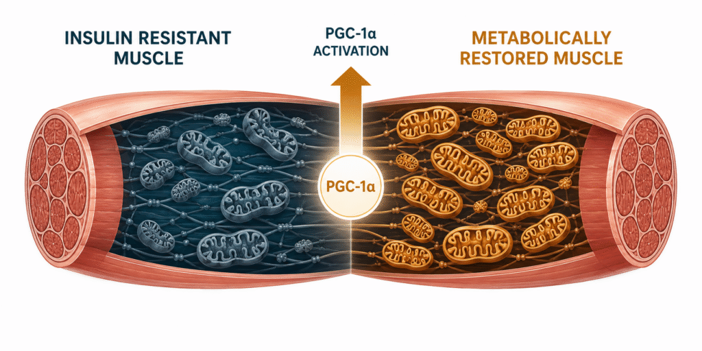
Mitochondria are the organelles responsible for producing ATP — the energy currency that powers every biological process in the body. In metabolically healthy tissue, they are dense, efficient, and capable of oxidizing both glucose and fat depending on what is available and what the cell demands. In insulin-resistant muscle, they are sparse, dysfunctional, and predominantly dependent on glucose — a fuel source that, in the context of chronic hyperinsulinemia, is simultaneously oversupplied and poorly handled. The result is a cellular energy crisis that manifests as fatigue, brain fog, poor exercise tolerance, and the progressive deterioration of metabolic function that characterizes the insulin resistance trajectory.
Understanding how mitochondrial density is lost, how it is rebuilt, and what that rebuilding looks like clinically changes what practitioners look for and what patients should expect from a metabolic recovery protocol.
What you will learn: What mitochondria are and why their density matters for insulin resistance | The mechanisms by which insulin resistance impairs mitochondrial function | How specific interventions rebuild mitochondrial density | What the clinical signals of improving mitochondrial function look like in practice | Why mitochondrial recovery is a convergent outcome of the full metabolic protocol rather than an isolated intervention
What Mitochondria Are and Why Density Matters
Every cell in the body — with the exception of mature red blood cells — contains mitochondria. In skeletal muscle, which is the metabolically most active tissue in the body and the site of 75 to 85 percent of postprandial glucose disposal, mitochondrial density determines the cell’s capacity to produce ATP aerobically from both glucose and fat. High mitochondrial density means the cell can generate energy efficiently across a wide range of fuel availability and metabolic demand. Low mitochondrial density means the cell is forced into less efficient metabolic pathways — primarily anaerobic glycolysis — that produce ATP faster but with greater metabolic cost and without the ability to oxidize fat effectively.
This distinction has direct clinical consequences. A patient with high mitochondrial density in skeletal muscle can oxidize fat at rest and at moderate exercise intensities, maintains stable blood glucose between meals because muscle can draw on fat rather than demanding glucose, and recovers quickly from physical exertion because ATP production capacity is robust. A patient with low mitochondrial density cannot oxidize fat effectively, depends heavily on glucose even at rest, experiences energy crashes when glucose availability fluctuates, and accumulates intramyocellular lipid — fat droplets within muscle fibers — that directly impairs insulin signaling through diacylglycerol-mediated PKC activation.
Mitochondrial density is not fixed. It responds directly to the demands placed on it. Use it and it grows. Underload it and it atrophies. The cellular machinery for creating new mitochondria — mitochondrial biogenesis — is one of the most trainable biological systems in the human body, and it responds to interventions that are entirely within the patient’s control.
How Insulin Resistance Impairs Mitochondrial Function
The relationship between insulin resistance and mitochondrial dysfunction is bidirectional — each condition drives and deepens the other through specific mechanisms that, once understood, explain why metabolic deterioration tends to accelerate rather than plateau once it begins.
Chronically elevated insulin, as detailed in the post on insulin resistance, impairs mitochondrial function through several converging pathways. Hyperinsulinemia suppresses fat oxidation by maintaining malonyl-CoA at levels that inhibit carnitine palmitoyltransferase I — the enzyme that transports fatty acids into the mitochondrial matrix for oxidation. When fat cannot enter the mitochondria, it accumulates as intramyocellular lipid. These lipid intermediates — particularly diacylglycerol and ceramides — activate serine kinases that phosphorylate IRS-1, converting the insulin receptor signaling cascade from active to inhibited. The result is a self-reinforcing loop: insulin resistance impairs fat oxidation, impaired fat oxidation generates lipid intermediates, lipid intermediates deepen insulin resistance.
Chronic hyperinsulinemia also suppresses PGC-1α — peroxisome proliferator-activated receptor gamma coactivator 1-alpha — the master transcriptional regulator of mitochondrial biogenesis. When insulin is chronically elevated, the cell receives a signal of nutrient abundance that reduces the adaptive stimulus for mitochondrial expansion. The body does not invest in building more energy production infrastructure when it believes it already has more fuel than it can handle.
The chronic low-grade inflammation that accompanies insulin resistance adds a further layer of mitochondrial damage. Inflammatory cytokines — particularly TNF-α and IL-6 from dysfunctional adipose tissue — generate reactive oxygen species that damage mitochondrial membranes, impair electron transport chain efficiency, and accelerate mitochondrial protein turnover faster than the biogenesis machinery can replace them. As covered in the post on skeletal muscle and metabolic health, this inflammatory-mitochondrial axis is one of the central mechanisms connecting visceral fat accumulation to the progressive metabolic deterioration observed in insulin-resistant patients.
How Mitochondrial Density Is Rebuilt: The Three Primary Mechanisms
Zone 2 Training and PGC-1α Activation
The most potent stimulus for mitochondrial biogenesis in skeletal muscle is sustained aerobic exercise at moderate intensity — what exercise physiologists call Zone 2. At this intensity, mitochondria are working near their oxidative capacity without being overwhelmed by lactate accumulation. The cellular energy deficit generated by sustained aerobic demand activates AMPK — AMP-activated protein kinase — which phosphorylates and activates PGC-1α. PGC-1α then drives the transcription of nuclear-encoded mitochondrial proteins, stimulates mitochondrial DNA replication, and initiates the coordinated expansion of the mitochondrial network within the muscle fiber.
This is the cellular mechanism behind the clinical observation that consistent Zone 2 walking produces qualitatively different metabolic adaptations than high-intensity training. As covered in detail in the post on zone 2 training, the intensity specificity matters: PGC-1α activation through the mitochondrial stress pathway is most robustly triggered at Zone 2 intensity, where the demand is sustained and the mitochondria are challenged without the alternative signaling pathways activated at higher intensities dominating the adaptive response.
Resistance Training and Mitochondrial Quality
Resistance training contributes to mitochondrial health through a distinct but complementary mechanism. Mechanical loading activates mTOR signaling and stimulates mitophagy — the selective degradation of damaged or dysfunctional mitochondria — alongside the biogenesis of new ones. The net effect is an improvement in mitochondrial quality: the dysfunctional mitochondria that are generating reactive oxygen species and impairing insulin signaling are cleared, and new, more efficient mitochondria replace them.
Resistance training also increases the total volume of skeletal muscle tissue — the cellular compartment in which mitochondria reside. More muscle mass means more mitochondrial housing capacity. This structural expansion amplifies the effect of Zone 2 training on total mitochondrial output, which is one of the primary reasons the combination of resistance training and Zone 2 walking produces superior metabolic outcomes to either modality alone. The interaction between resistance training, muscle mass, and metabolic function is examined in the post on resistance training and metabolism.
Dietary Ketosis, Fasting, and Metabolic Switching
Periods of low insulin — achieved through carbohydrate restriction, time-restricted eating, or intermittent fasting — create a metabolic state in which the cell must rely on fat oxidation for ATP production. This metabolic demand activates the same AMPK-PGC-1α pathway that Zone 2 exercise engages, driving mitochondrial biogenesis through a nutritional rather than exercise stimulus. The two mechanisms are additive: patients who combine consistent Zone 2 training with a dietary approach that produces regular low-insulin periods generate a stronger and more sustained biogenesis stimulus than either intervention alone produces.
The animal-based, low-refined-carbohydrate nutritional framework also removes the chronic hyperinsulinemia that was suppressing PGC-1α in the first place — allowing the biogenesis machinery to operate without the hormonal brake that insulin excess had been applying.

Clinical Perspective: What Improving Mitochondrial Density Looks Like in Practice
In clinical practice, mitochondrial density cannot be measured directly without a muscle biopsy — a tool that belongs in research settings, not routine clinical care. What can be observed is the functional fingerprint of improving mitochondrial capacity: a consistent pattern of changes across symptoms, exercise physiology, and metabolic markers that together tell a coherent physiological story.
The earliest signal is almost always subjective and precedes any laboratory change. Patients report that they can sustain the same pace of walking with less perceived effort and recover more quickly afterward. Many notice more stable energy throughout the day — the afternoon fatigue that had become normalized begins to lift. Mental clarity improves. Cravings reduce. These are functional signs that the cell is producing ATP more efficiently and accessing fat as a fuel source more readily between meals — the early expression of restored metabolic flexibility.
Objectively, I look for improvements in resting heart rate and heart rate variability — both markers of improved autonomic balance and cardiovascular efficiency that reflect underlying mitochondrial adaptation. Aerobic capacity at a given workload improves measurably: the patient reaches Zone 2 at a faster pace as fitness develops, which is itself a reliable proxy for mitochondrial density improvement. As covered in the post on recovery and metabolic adaptation, heart rate variability is the most sensitive available clinical marker of the autonomic recovery that mitochondrial adaptation supports.
Metabolically, the picture that emerges over eight to twelve weeks of consistent intervention includes falling fasting insulin, improving TG/HDL ratio, rising HDL, normalizing liver enzymes in patients with fatty liver, and more stable postprandial glucose regulation. None of these markers proves increased mitochondrial density independently. But their convergence — particularly the combination of improved exercise tolerance, stable energy, and improving upstream metabolic markers — tells a story that is consistent with the mitochondrial adaptation the interventions are designed to produce.
The important clinical caveat is that I do not attribute these improvements solely to mitochondrial biogenesis. Exercise, nutrition, sleep quality, reduction of ectopic fat, and resolution of chronic hyperinsulinemia all interact simultaneously in a well-implemented protocol. Improved mitochondrial function is one critical adaptation within a broader restoration of metabolic health — it is the engine upgrade, but the engine operates within a vehicle that every other intervention is also improving at the same time. The convergence of markers and symptoms is what builds clinical confidence that the biology is moving in the right direction.
The patients who show the slowest mitochondrial recovery are predictable: those combining chronic cardio stress with inadequate sleep, those whose training volume exceeds recovery capacity and suppresses PGC-1α through cortisol excess, and those whose dietary pattern keeps insulin elevated throughout the day — blocking fat oxidation and maintaining the hormonal environment that suppresses mitochondrial biogenesis regardless of training stimulus. As covered in the posts on myokines and muscle as an endocrine organ, the full mitochondrial benefit of exercise requires the hormonal and recovery environment to support the adaptation — not just the training stimulus to initiate it.
Mitochondrial Density, Sarcopenia, and the Aging Trajectory
The decline of mitochondrial density with aging is one of the most consequential and least discussed biological processes in metabolic medicine. Mitochondrial function declines measurably from the fourth decade onward through a combination of reduced physical demand, accumulating mitochondrial DNA damage from reactive oxygen species, declining anabolic hormone support for mitochondrial protein synthesis, and the progressive reduction of PGC-1α expression that accompanies both aging and metabolic dysfunction.
This mitochondrial decline runs in parallel with the muscle loss that characterizes sarcopenia — and the two processes amplify each other. Fewer mitochondria mean less capacity to oxidize fat within muscle fibers, which accelerates intramyocellular lipid accumulation, which deepens insulin resistance within the remaining muscle tissue, which reduces the anabolic signaling that would otherwise maintain muscle mass. The loss of muscle mass reduces the total mitochondrial housing capacity, which reduces total mitochondrial output, which further impairs the metabolic function of the tissue that remains.
This trajectory is not inevitable. The same interventions that drive mitochondrial biogenesis in younger patients — Zone 2 training, resistance loading, adequate protein, low-insulin nutritional periods, and sleep optimization — produce measurable mitochondrial adaptation in older adults. The rate of adaptation is slower and the anabolic resistance means a higher training and protein stimulus is required to achieve the same response, but the machinery responds. Mitochondrial biogenesis does not have an age cutoff.
A Note on Uncertainty
The clinical translation of mitochondrial density research contains important limitations. Most of the mechanistic evidence for PGC-1α activation, mitochondrial biogenesis thresholds, and the precise dose-response relationships between Zone 2 training volume and mitochondrial density comes from research in trained athletes and young healthy subjects. Direct translation to deconditioned, insulin-resistant, and older patient populations — while directionally supported — is less precisely characterized.
Indirect clinical markers of mitochondrial function, including heart rate variability, resting heart rate, and exercise tolerance, are proxies rather than direct measurements. Individual variation in mitochondrial adaptation rate is substantial and depends on genetic factors, hormonal status, sleep quality, and baseline metabolic health. What the evidence consistently supports is the directional framework: the interventions described in this post drive mitochondrial biogenesis through well-characterized pathways, and the functional improvements that follow are measurable, clinically meaningful, and reproducible across the patient populations in which they have been studied.

The Animal-Based Protocol for Insulin Resistance and Type 2 Diabetes
A Functional Medicine Framework
People Also Ask
What is mitochondrial density and why does it matter?
Mitochondrial density refers to the number and functional capacity of mitochondria within a cell, particularly in skeletal muscle. Higher mitochondrial density means greater capacity for aerobic ATP production, fat oxidation, and metabolic flexibility. Lower density forces cells into less efficient metabolic pathways, impairs fat oxidation, and contributes to insulin resistance through intramyocellular lipid accumulation.
How does mitochondrial dysfunction cause insulin resistance?
Mitochondrial dysfunction impairs fat oxidation within muscle fibers, leading to accumulation of lipid intermediates — diacylglycerol and ceramides — that activate serine kinases blocking the insulin receptor signaling cascade. This is one of the primary cellular mechanisms by which reduced mitochondrial capacity directly causes peripheral insulin resistance independently of dietary carbohydrate intake.
What is the best exercise for mitochondrial biogenesis?
Zone 2 training — sustained aerobic exercise at 60 to 70 percent of maximum heart rate — produces the most robust PGC-1α activation and mitochondrial biogenesis stimulus. Resistance training improves mitochondrial quality through mitophagy and structural muscle expansion. The combination of both modalities produces superior mitochondrial adaptation to either alone.
How long does it take to improve mitochondrial density?
Early functional improvements — better energy, improved exercise tolerance, more stable glucose — typically appear within two to four weeks of consistent Zone 2 training. Meaningful structural mitochondrial adaptation, measurable as improved aerobic capacity and fat oxidation at a given workload, develops over eight to twelve weeks. Full mitochondrial density restoration in significantly insulin-resistant patients may require months of consistent intervention.
Can mitochondrial density be measured clinically?
Direct measurement requires muscle biopsy — a research tool not available in routine clinical care. Clinically, mitochondrial density improvement is inferred from a convergent pattern: improving exercise tolerance at a given workload, rising heart rate variability, falling resting heart rate, more stable energy and reduced fatigue, and improving metabolic markers including fasting insulin, TG/HDL ratio, and postprandial glucose regulation.
Does aging irreversibly reduce mitochondrial density?
No. Mitochondrial biogenesis responds to training and nutritional stimuli at every age. The rate of adaptation is slower in older adults and the stimulus required is higher due to anabolic resistance, but the machinery remains responsive. Zone 2 training and resistance loading produce measurable mitochondrial adaptation in older patients — the trajectory of age-related mitochondrial decline is modifiable, not fixed.
Why does insulin resistance impair fat oxidation?
Chronic hyperinsulinemia maintains malonyl-CoA at levels that inhibit carnitine palmitoyltransferase I — the enzyme responsible for transporting fatty acids into the mitochondrial matrix for oxidation. When this transport is inhibited, fat cannot be oxidized efficiently regardless of its availability, and cells default to glucose as their primary fuel source even when glucose supply is inadequate or insulin signaling is impaired.
About the Author
Morteza Ariana is a State-Certified Functional Nutritionist based in Germany, specializing in insulin resistance, type 2 diabetes, and root-cause metabolic restoration. He holds advanced training in systems-based physiology and has worked with patients across the U.S. and Europe for over 10 years.
His clinical framework is built around a core principle that mainstream medicine consistently overlooks: chronically elevated insulin — not blood glucose — is the earliest and most actionable driver of metabolic disease. That conviction was shaped in part by his own experience with hyperinsulinemia in 2016, and deepened through a decade of clinical practice and the study of leading researchers in metabolic medicine including Benjamin Bikman, Joseph Kraft, Gerald Reaven, Jason Fung, and Stephen Phinney.
His work focuses on identifying and correcting the upstream metabolic signals — insulin load, liver-gut axis dysfunction, circadian misalignment, and micronutrient gaps — that standard screening misses entirely. Patient outcomes are documented, anonymized, and published on this site.
If this resonates, the next step is clarity
The Metabolic Restoration Blueprint is a structured 12-week framework designed to correct upstream metabolic drivers — not just manage symptoms.
Scientific References
Holloszy JO. Exercise-induced increase in muscle insulin sensitivity. J Appl Physiol. 2005;99(1):338–343. 🔗 https://pubmed.ncbi.nlm.nih.gov/16036907/
Petersen KF, Befroy D, Dufour S, et al. Mitochondrial dysfunction in the elderly: possible role in insulin resistance. Science. 2003;300(5622):1140–1142. 🔗 https://pubmed.ncbi.nlm.nih.gov/12750520/
Finck BN, Kelly DP. PGC-1 coactivators: inducible regulators of energy metabolism in health and disease. J Clin Invest. 2006;116(3):615–622. 🔗 https://pubmed.ncbi.nlm.nih.gov/16511594/
Holloszy JO, Coyle EF. Adaptations of skeletal muscle to endurance exercise and their metabolic consequences. J Appl Physiol. 1984;56(4):831–838. 🔗 https://pubmed.ncbi.nlm.nih.gov/6373687/
Menshikova EV, Ritov VB, Fairfull L, et al. Effects of exercise on mitochondrial content and function in aging human skeletal muscle. J Gerontol A Biol Sci Med Sci. 2006;61(6):534–540. 🔗 https://pubmed.ncbi.nlm.nih.gov/16799133/
Kelley DE, He J, Menshikova EV, Ritov VB. Dysfunction of mitochondria in human skeletal muscle in type 2 diabetes. Diabetes. 2002;51(10):2944–2950. 🔗 https://pubmed.ncbi.nlm.nih.gov/12351431/
Morino K, Petersen KF, Shulman GI. Molecular mechanisms of insulin resistance in humans and their potential links with mitochondrial dysfunction. Diabetes. 2006;55(Suppl 2):S9–S15. 🔗 https://pubmed.ncbi.nlm.nih.gov/17130651/
Goodpaster BH, Sparks LM. Metabolic flexibility in health and disease. Cell Metab. 2017;25(5):1027–1036. 🔗 https://pubmed.ncbi.nlm.nih.gov/28467930/
Irrcher I, Adhihetty PJ, Joseph AM, et al. Regulation of mitochondrial biogenesis in muscle by endurance exercise. Sports Med. 2003;33(11):783–793. 🔗 https://pubmed.ncbi.nlm.nih.gov/12959619/
Richter EA, Hargreaves M. Exercise, GLUT4, and skeletal muscle glucose uptake. Physiol Rev. 2013;93(3):993–1017. 🔗 https://pubmed.ncbi.nlm.nih.gov/23899560/
Lanza IR, Sreekumaran Nair K. Muscle mitochondrial changes with aging and exercise. Am J Clin Nutr. 2009;89(1):467S–471S. 🔗 https://pubmed.ncbi.nlm.nih.gov/19118214/
Pedersen BK, Febbraio MA. Muscles, exercise and obesity: skeletal muscle as a secretory organ. Nat Rev Endocrinol. 2012;8(8):457–465. 🔗 https://pubmed.ncbi.nlm.nih.gov/22473333/
Samuel VT, Shulman GI. Mechanisms for insulin resistance: common threads and missing links. Cell. 2012;148(5):852–871. 🔗 https://pubmed.ncbi.nlm.nih.gov/22385956/
Hood DA, Tryon LD, Carter HN, et al. Unravelling the mechanisms regulating muscle mitochondrial biogenesis. Biochem J. 2016;473(15):2295–2314. 🔗 https://pubmed.ncbi.nlm.nih.gov/27470593/