
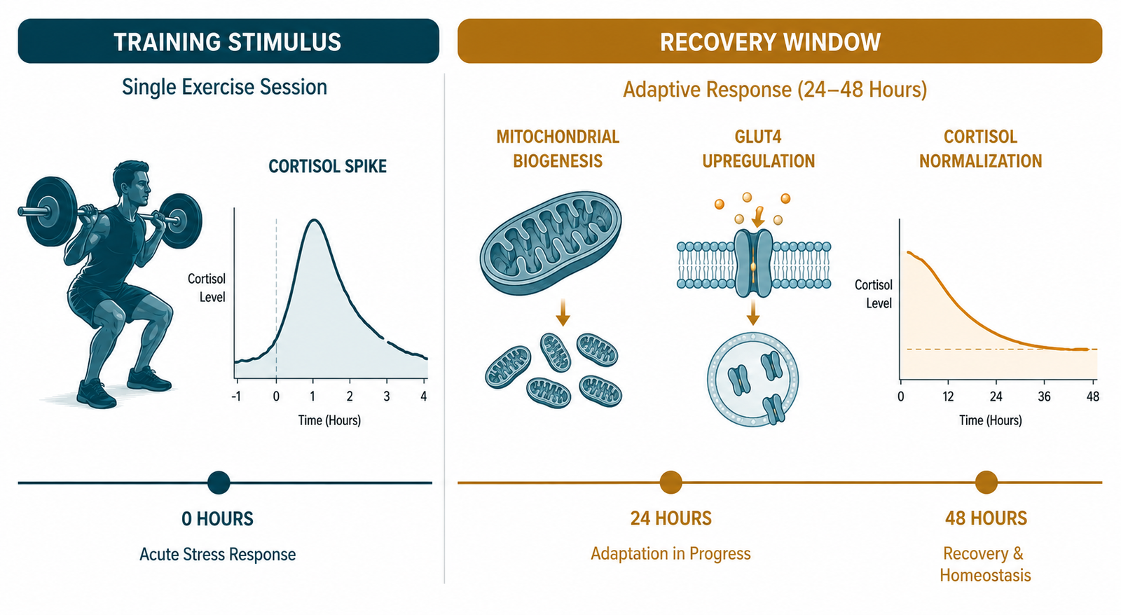
Recovery and metabolic adaptation are two phases of the same process — the training session provides the stimulus, but adaptation only completes when recovery conditions are met.
There is a widespread assumption in health and fitness culture that progress is proportional to effort. Train harder, eat cleaner, push through the plateau. When results stall, the default prescription is more — more sessions, more restriction, more discipline. What this framework misses entirely is that the adaptation to training does not occur during exercise. It occurs during recovery. Exercise provides the stimulus. Recovery is when the body actually changes.
This distinction is not semantic. It has direct physiological consequences for insulin-resistant patients who are already carrying a significant allostatic load — the cumulative burden of metabolic dysfunction, chronic stress, poor sleep, and years of compensatory hyperinsulinemia. In these patients, recovery is not a passive rest period between workouts. It is an active biological process that determines whether the training stimulus produces adaptation or accumulates as additional stress on a system that is already struggling to cope.
Understanding recovery as a metabolic intervention in its own right — not an optional add-on to a training program — changes what clinicians look for, what they prioritize, and why progress so often stalls in patients who appear to be doing everything right.
What you will learn: Why adaptation occurs during recovery rather than during exercise | The clinical pattern that identifies insufficient recovery as the bottleneck | What the markers and symptoms of impaired recovery look like | How autonomic balance, sleep, and protein intake determine recovery quality | What a practical recovery-first protocol looks like for a metabolically compromised patient
The Physiology of Adaptation: What Recovery Actually Does
When skeletal muscle contracts under load, several things happen simultaneously. Muscle fibers sustain microdamage. Glycogen stores are depleted. Reactive oxygen species are generated. Cortisol rises. The immune system mounts a localized inflammatory response. These are not side effects of training — they are the signals that initiate adaptation.
In the hours and days following a training session, provided recovery conditions are adequate, a coordinated repair process unfolds. Satellite cells — muscle stem cells — are activated and begin rebuilding damaged fibers with slightly greater diameter and strength. Mitochondrial biogenesis proceeds through PGC-1α activation, increasing mitochondrial density and fat oxidation capacity. GLUT4 transporter expression is upregulated, improving insulin-independent glucose disposal. Inflammatory cytokines resolve. Cortisol returns to baseline. The autonomic nervous system shifts from sympathetic dominance back toward parasympathetic tone, allowing heart rate variability to recover and the hormonal environment to normalize.
This entire process — the actual physiological adaptation — requires sleep, adequate protein, sufficient total energy, low background inflammatory load, and a cumulative stress environment that leaves enough biological resources for repair. Remove any of those inputs, and the adaptation is incomplete. The training stimulus accumulates as unresolved stress rather than resolved improvement. The patient trains consistently, adheres to their nutrition protocol, and makes no progress — because the recovery phase that would translate stimulus into adaptation never fully completes.
When Recovery Is the Bottleneck: The Clinical Pattern
Recovery insufficiency rarely presents as a single dramatic symptom. It presents as a pattern — a cluster of findings that, taken together, point to a system operating in a state of chronic sympathetic activation rather than healthy adaptation.
The symptom pattern is consistent across patients: persistent fatigue despite adequate sleep duration, declining motivation to exercise despite genuine commitment, muscle soreness lasting longer than expected after sessions, reduced training performance over time rather than improvement, brain fog, irritability, poor stress tolerance, and difficulty losing fat despite good dietary adherence. Many patients also report waking unrefreshed or experiencing early morning awakenings — the 3 to 4 a.m. cortisol-driven arousal that reflects dysregulated HPA axis activity rather than adequate sleep architecture.
The physiological markers tell a parallel story. Resting heart rate remains elevated rather than declining with fitness improvement. Heart rate variability — the most sensitive available marker of autonomic recovery — is reduced in patients with access to measurement tools. Fasting glucose is unexpectedly elevated in the morning, reflecting nocturnal gluconeogenesis driven by cortisol excess rather than dietary intake. Fasting insulin remains elevated despite lifestyle improvements, because the chronic sympathetic activation and inflammatory load are impairing insulin signaling independently of the nutritional changes being made. Inflammatory markers including hsCRP remain mildly elevated. In some patients, liver enzymes — particularly GGT — reflect ongoing metabolic stress, though these always require interpretation in full clinical context.
What makes recovery insufficiency particularly difficult to identify is that it mimics insufficient effort. The patient looks like someone who needs to work harder. The markers look like someone whose protocol is not aggressive enough. The correct diagnosis — that the system is overtaxed and needs less stimulus and more recovery — runs directly counter to the cultural default of pushing through plateaus.

The Autonomic Dimension: Sympathetic Dominance and Metabolic Consequences
One of the most important and least discussed aspects of recovery insufficiency is its effect on autonomic balance. The autonomic nervous system operates through two complementary branches: the sympathetic system, which mobilizes energy and resources for effort, and the parasympathetic system, which governs repair, digestion, immune regulation, and metabolic restoration. Healthy adaptation to training requires cycling between these states — sympathetic activation during exercise, parasympathetic dominance during recovery.
When training volume consistently exceeds recovery capacity, this cycling breaks down. The system becomes stuck in sympathetic dominance — a state of chronic low-level activation that generates metabolic consequences beyond simple fatigue. Chronically elevated sympathetic tone increases cortisol and catecholamine output, raising fasting glucose through hepatic gluconeogenesis. It impairs insulin-stimulated glucose uptake in peripheral tissues through adrenergic receptor activation. It suppresses growth hormone secretion — which occurs predominantly during deep slow-wave sleep — reducing the anabolic stimulus that would otherwise drive muscle protein synthesis overnight. It elevates resting heart rate, reduces heart rate variability, and maintains the body in a catabolic rather than anabolic hormonal environment.
This is the physiological substrate of the “wired but tired” presentation that characterizes chronic stress and recovery insufficiency: the sympathetic system is active, cortisol is elevated, the patient cannot wind down at night, and yet the tissues are depleted rather than energized. As covered in the post on chronic cardio stress, this pattern is particularly common in patients combining high exercise volume with inadequate sleep and chronic occupational or psychological stress.
Sleep as the Primary Recovery Intervention
Of all the variables that determine recovery quality, sleep is the most powerful and the most frequently under-prioritized. The metabolic consequences of insufficient sleep are profound, well-documented, and clinically underestimated.
During slow-wave sleep — the deepest stage of non-REM sleep — growth hormone is secreted in its largest daily pulse. This growth hormone surge drives muscle protein synthesis, supports fat oxidation, and maintains the anabolic hormonal environment that allows training adaptations to consolidate. A patient who trains consistently but sleeps six hours or less is chronically suppressing the primary hormonal driver of the adaptations their training is intended to produce.
Sleep deprivation also elevates cortisol, reduces insulin sensitivity through mechanisms independent of diet and exercise, increases ghrelin and reduces leptin — shifting appetite regulation toward overconsumption — and impairs the glucose metabolism that determines whether carbohydrate substrate is stored as glycogen or diverted to fat. A single night of poor sleep measurably reduces insulin sensitivity the following day. Chronic sleep insufficiency produces a persistent metabolic impairment that no training protocol can fully overcome.
For insulin-resistant patients — whose metabolic system is already under significant stress — sleep optimization is not a lifestyle preference. It is a clinical priority that sits alongside protein intake and resistance training in the hierarchy of metabolic interventions. The post on skeletal muscle and metabolic health covers the interaction between sleep disruption, cortisol, and muscle catabolism in the context of the broader metabolic picture.
Clinical Perspective: What I See in Practice
Recovery is one of the most commonly overlooked bottlenecks in clinical practice. I encounter it regularly in patients who are genuinely committed — adequate protein, consistent resistance training and metabolism protocols in place, zone 2 training established, diet clean — but whose progress has plateaued in a way that does not respond to further dietary refinement.
Before I conclude that recovery is the limiting variable, I rule out other contributors: inadequate protein intake, insufficient total energy, thyroid dysfunction, nutrient deficiencies including vitamin D and magnesium, and excessive training volume. If those are reasonably addressed and the plateau persists, recovery moves to the top of the list.
The pattern I look for is consistent. Patients describe persistent fatigue despite sleeping adequately, soreness that takes longer than expected to resolve, declining training performance rather than improvement, brain fog that does not clear across the day, and a generalized sense of effort without return. When I look at their markers, fasting insulin remains elevated despite dietary compliance, hsCRP is mildly but persistently elevated, and fasting glucose is higher in the morning than expected — the fingerprint of nocturnal cortisol-driven gluconeogenesis.
The intervention that most reliably restores progress is not more training. It is replacing some training volume with structured recovery: prioritizing sleep as a first-order intervention, introducing rest days with deliberate low-intensity movement rather than additional sessions, ensuring protein is distributed adequately across meals to support overnight muscle protein synthesis, and addressing the psychological and occupational stress load that is contributing to the sympathetic dominance pattern.
What I consistently observe is that patients who accept this reframe — who shift from “I need to push harder” to “I need to recover better” — begin making progress again within four to eight weeks. Not because their training has become more aggressive, but because the adaptation that their training was already generating finally has the conditions it needs to complete.
The key message I return to in every conversation about recovery: exercise is the stimulus. Recovery is when the body actually changes. Without the second half of that equation, the first half is biological noise.
Protein, Energy Availability, and Overnight Repair
Recovery quality is not determined by rest alone. It requires adequate substrate — specifically protein and total energy — to execute the repair processes that training initiates.
Muscle protein synthesis is a leucine-threshold phenomenon: each meal needs to deliver sufficient leucine to trigger the mTOR signaling cascade that initiates rebuilding. A patient who trains consistently but distributes protein poorly across the day — eating little at breakfast, a moderate lunch, and a large protein load at dinner — is providing the overnight repair stimulus only once, rather than maintaining the conditions for synthesis across the full recovery window. The practical target of 1.6 grams of protein per kilogram of ideal body weight, distributed across meals rather than concentrated in one, reflects this biology directly.
Total energy availability matters independently of protein. Chronic caloric restriction — common in patients who are simultaneously trying to lose fat and build muscle — creates an energy deficit that the body resolves by reducing muscle protein synthesis rates and downregulating thermogenesis. The result is impaired recovery alongside impaired metabolic adaptation. For insulin-resistant patients this creates a clinical tension: sufficient caloric restriction to reduce insulin load, but not so severe that energy availability impairs the recovery that training requires. Protein-adequate, moderate caloric restriction — rather than aggressive deficit — is the balance that most consistently supports both goals simultaneously.
The Role of Myokines in Recovery
The recovery period is not only when structural repair occurs. It is also when the myokine signals generated during exercise exert their systemic effects. As covered in the posts on muscle as an endocrine organ and myokines, muscle-derived IL-6, irisin, BDNF, and FGF-21 are released during contraction and act on the liver, adipose tissue, brain, and pancreas in the hours following exercise. These signals require a low-inflammation, parasympathetically balanced recovery environment to exert their full effects. When the background inflammatory load is elevated — from overtraining, poor sleep, or chronic stress — myokine signaling is partially drowned out. The metabolic communication that contraction is attempting to broadcast cannot be received clearly.
This is one reason why recovery optimization amplifies the benefit of training rather than simply preserving it. Better recovery does not just prevent overtraining — it improves the signal-to-noise ratio of the biological communication that makes training metabolically productive in the first place.
Sarcopenia and the Recovery Deficit
The interaction between impaired recovery and sarcopenia deserves specific attention. Sarcopenia — the progressive loss of skeletal muscle mass and function — is driven in part by a reduced anabolic response to training stimulus that develops with aging and metabolic dysfunction. Older and insulin-resistant patients require more recovery per training session to achieve the same adaptive response as younger, metabolically healthy individuals, because anabolic resistance means the mTOR signaling response to protein and mechanical loading is blunted.
This has a direct practical implication: the recovery requirements of a 55-year-old insulin-resistant patient are not the same as those of a 30-year-old healthy athlete. Training programs designed for the latter — high frequency, high volume, short recovery windows — are not appropriate for the former. Matching recovery time to the patient’s actual anabolic response capacity, rather than to an idealized training template, is one of the most important individualizations in a metabolic recovery protocol.

The Animal-Based Protocol for Insulin Resistance and Type 2 Diabetes
A Functional Medicine Framework
A Note on Uncertainty
The recovery science, while directionally consistent, contains important limitations. Heart rate variability as a recovery marker is promising but lacks standardized clinical cutoffs. The optimal protein distribution for overnight muscle protein synthesis in insulin-resistant patients specifically is still being refined. The precise contribution of psychological stress versus training stress to HPA axis dysregulation in individual patients is difficult to disentangle. And the recovery requirements of metabolically compromised patients have been studied far less systematically than those of athletic populations. What the evidence consistently supports is the directional framework: recovery is an active biological process that requires sleep, protein, energy availability, and low background stress to complete — and optimizing it produces metabolic improvements that additional training volume does not.
People Also Ask
Why is recovery important for metabolic health?
Recovery is when the physiological adaptations to training actually occur — muscle protein synthesis, mitochondrial biogenesis, GLUT4 upregulation, cortisol normalization, and autonomic rebalancing. Without adequate recovery, training stimulus accumulates as unresolved stress rather than completed adaptation, and metabolic progress stalls regardless of training quality.
What are the signs of insufficient recovery?
Persistent fatigue despite adequate sleep, declining training performance, prolonged muscle soreness, brain fog, irritability, poor stress tolerance, elevated resting heart rate, reduced heart rate variability, unexpectedly high morning fasting glucose, and difficulty losing fat despite dietary adherence — taken together, these form the clinical pattern of recovery insufficiency.
How does sleep affect metabolic adaptation?
Slow-wave sleep drives the largest daily pulse of growth hormone, which is essential for muscle protein synthesis and fat oxidation. Sleep deprivation elevates cortisol, reduces insulin sensitivity, impairs glucose metabolism, and suppresses the anabolic hormonal environment that training adaptations require. For insulin-resistant patients, sleep optimization is a primary metabolic intervention, not a lifestyle preference.
Can you overtrain if you have insulin resistance?
Yes — and the threshold for overtraining is lower in metabolically compromised patients because their recovery capacity is already reduced by chronic inflammation, HPA axis stress, poor sleep, and impaired anabolic signaling. High training volumes that would be appropriate for a healthy athlete can produce chronic stress responses in insulin-resistant patients that deepen rather than improve metabolic dysfunction.
What is heart rate variability and why does it matter for recovery?
Heart rate variability is the variation in time between successive heartbeats, reflecting the balance between sympathetic and parasympathetic nervous system activity. Higher HRV indicates better autonomic recovery and parasympathetic tone. Reduced HRV is a reliable early marker of insufficient recovery, overtraining, and chronic stress. It is the most sensitive available tool for monitoring recovery status in patients with access to measurement.
How much protein is needed for recovery from resistance training?
A minimum of 1.6 grams per kilogram of ideal body weight per day, distributed across meals to ensure leucine threshold is met at each sitting. Older patients and those with anabolic resistance may require up to 2.0 grams per kilogram. Protein concentration in a single evening meal does not substitute for distribution across the recovery window.
How long does metabolic adaptation take?
Meaningful mitochondrial adaptation and GLUT4 upregulation begin within two to four weeks of consistent training with adequate recovery. Structural muscle adaptation takes eight to twelve weeks. Full metabolic flexibility restoration — the capacity to shift efficiently between glucose and fat — develops over months of consistent training, adequate protein, appropriate meal spacing, and sleep optimization working together.
About the Author
Morteza Ariana is a State-Certified Functional Nutritionist based in Germany, specializing in insulin resistance, type 2 diabetes, and root-cause metabolic restoration. He holds advanced training in systems-based physiology and has worked with patients across the U.S. and Europe for over 10 years.
His clinical framework is built around a core principle that mainstream medicine consistently overlooks: chronically elevated insulin — not blood glucose — is the earliest and most actionable driver of metabolic disease. That conviction was shaped in part by his own experience with hyperinsulinemia in 2016, and deepened through a decade of clinical practice and the study of leading researchers in metabolic medicine including Benjamin Bikman, Joseph Kraft, Gerald Reaven, Jason Fung, and Stephen Phinney.
His work focuses on identifying and correcting the upstream metabolic signals — insulin load, liver-gut axis dysfunction, circadian misalignment, and micronutrient gaps — that standard screening misses entirely. Patient outcomes are documented, anonymized, and published on this site.
If this resonates, the next step is clarity
The Metabolic Restoration Blueprint is a structured 12-week framework designed to correct upstream metabolic drivers — not just manage symptoms.
Scientific References
Volpi E, Mittendorfer B, Rasmussen BB, Wolfe RR. The response of muscle protein anabolism to combined hyperaminoacidemia and glucose-induced hyperinsulinemia is impaired in the elderly. J Clin Endocrinol Metab. 2000;85(12):4481–4490. 🔗 https://pubmed.ncbi.nlm.nih.gov/11134099/
Kreher JB, Schwartz JB. Overtraining syndrome: a practical guide. Sports Health. 2012;4(2):128–138. 🔗 https://pubmed.ncbi.nlm.nih.gov/23016079/
Meeusen R, Duclos M, Foster C, et al. Prevention, diagnosis, and treatment of the overtraining syndrome. Med Sci Sports Exerc. 2013;45(1):186–205. 🔗 https://pubmed.ncbi.nlm.nih.gov/23247672/
Van Cauter E, Leproult R, Plat L. Age-related changes in slow wave sleep and REM sleep and relationship with growth hormone and cortisol levels in healthy men. JAMA. 2000;284(7):861–868. 🔗 https://pubmed.ncbi.nlm.nih.gov/10938176/
Leproult R, Van Cauter E. Role of sleep and sleep loss in hormonal release and metabolism. Endocr Dev. 2010;17:11–21. 🔗 https://pubmed.ncbi.nlm.nih.gov/19955752/
Spiegel K, Tasali E, Penev P, Van Cauter E. Sleep curtailment in healthy young men is associated with decreased leptin levels, elevated ghrelin levels, and increased hunger and appetite. Ann Intern Med. 2004;141(11):846–850. 🔗 https://pubmed.ncbi.nlm.nih.gov/15583226/
Moore DR, Robinson MJ, Fry JL, et al. Ingested protein dose response of muscle and albumin protein synthesis after resistance exercise in young men. Am J Clin Nutr. 2009;89(1):161–168. 🔗 https://pubmed.ncbi.nlm.nih.gov/19056590/
Morton RW, Murphy KT, McKellar SR, et al. A systematic review, meta-analysis and meta-regression of the effect of protein supplementation on resistance training-induced gains in muscle mass and strength. Br J Sports Med. 2018;52(6):376–384. 🔗 https://pubmed.ncbi.nlm.nih.gov/28698222/
Hawley JA, Lessard SJ. Exercise training-induced improvements in insulin action. Acta Physiol. 2008;192(1):127–135. 🔗 https://pubmed.ncbi.nlm.nih.gov/18171429/
Richter EA, Hargreaves M. Exercise, GLUT4, and skeletal muscle glucose uptake. Physiol Rev. 2013;93(3):993–1017. 🔗 https://pubmed.ncbi.nlm.nih.gov/23899560/
Pedersen BK, Febbraio MA. Muscles, exercise and obesity: skeletal muscle as a secretory organ. Nat Rev Endocrinol. 2012;8(8):457–465. 🔗 https://pubmed.ncbi.nlm.nih.gov/22473333/
Finck BN, Kelly DP. PGC-1 coactivators: inducible regulators of energy metabolism in health and disease. J Clin Invest. 2006;116(3):615–622. 🔗 https://pubmed.ncbi.nlm.nih.gov/16511594/
Cadegiani FA, Kater CE. Hypothalamic-pituitary-adrenal (HPA) axis functioning in overtraining syndrome. Endocr Connect. 2017;6(8):R167–R181. 🔗 https://pubmed.ncbi.nlm.nih.gov/29038160/
Hackney AC. Stress and the neuroendocrine system: the role of exercise as a stressor and modifier of stress. Expert Rev Endocrinol Metab. 2006;1(6):783–792. 🔗 https://pubmed.ncbi.nlm.nih.gov/30754144/